2ITP image
Entry Detail
PDB ID:
2ITP
Keywords:
Title:
Crystal structure of EGFR kinase domain G719S mutation in complex with AEE788
Biological Source:
Source Organism:
Host Organism:
PDB Version:
Deposition Date:
2006-05-25
Release Date:
2007-04-03
Method Details:
Experimental Method:
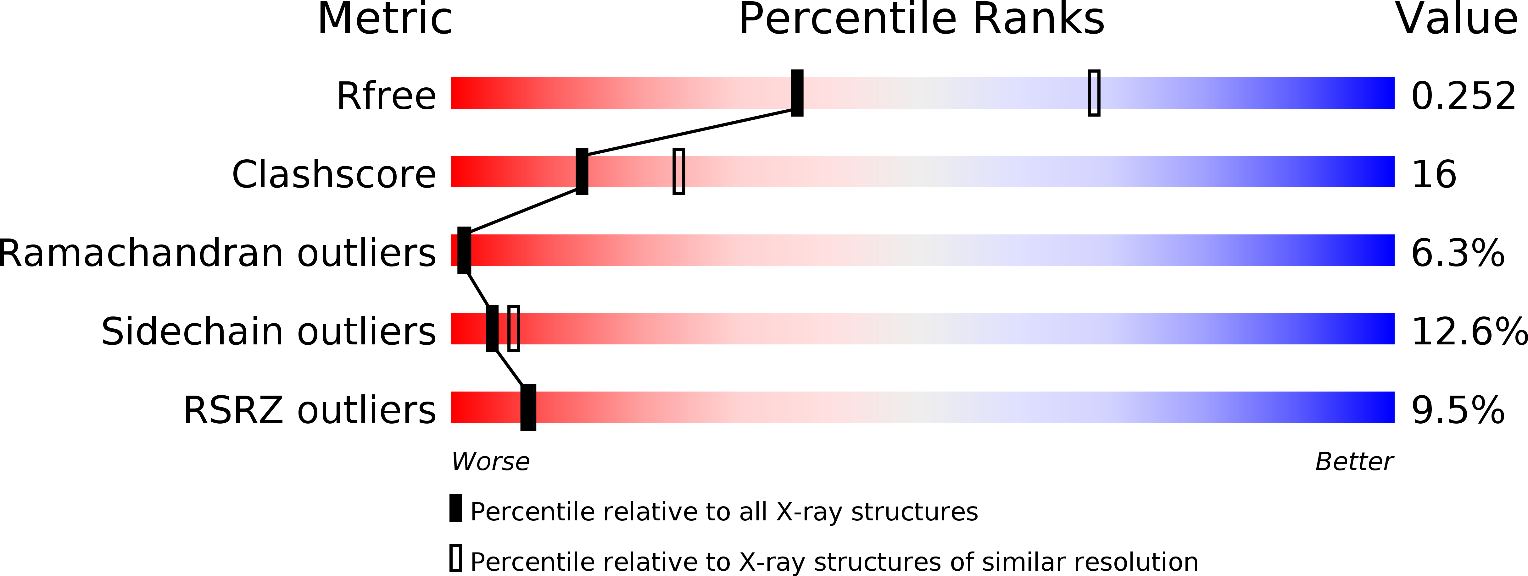
Resolution:
2.74 Å
R-Value Free:
0.25
R-Value Work:
0.19
R-Value Observed:
0.20
Space Group:
I 2 3
Macromolecular Entities
Polymer Type:polypeptide(L)
Description:EPIDERMAL GROWTH FACTOR RECEPTOR PRECURSOR
Mutations:YES
Chain IDs:A
Chain Length:327
Number of Molecules:1
Biological Source:HOMO SAPIENS
Ligand Molecules
Primary Citation
Structures of Lung Cancer-Derived Egfr Mutants and Inhibitor Complexes: Mechanism of Activation and Insights Into Differential Inhibitor Sensitivity
Cancer Cell 11 217 ? (2007)
PMID: 17349580 DOI: 10.1016/J.CCR.2006.12.017

Abstact

Mutations in the EGFR kinase are a cause of non-small-cell lung cancer. To understand their mechanism of activation and effects on drug binding, we studied the kinetics of the L858R and G719S mutants and determined their crystal structures with inhibitors including gefitinib, AEE788, and a staurosporine. We find that the mutations activate the kinase by disrupting autoinhibitory interactions, and that they accelerate catalysis as much as 50-fold in vitro. Structures of inhibitors in complex with both wild-type and mutant kinases reveal similar binding modes for gefitinib and AEE788, but a marked rotation of the staurosporine in the G719S mutant. Strikingly, direct binding measurements show that gefitinib binds 20-fold more tightly to the L858R mutant than to the wild-type enzyme.

Legend

Protein

Chemical

Disease

Primary Citation of related structures