2ISY image
Entry Detail
PDB ID:
2ISY
Keywords:
Title:
Crystal structure of the nickel-activated two-domain iron-dependent regulator (IdeR)
Biological Source:
PDB Version:
Deposition Date:
2006-10-18
Release Date:
2007-02-13
Method Details:
Experimental Method:
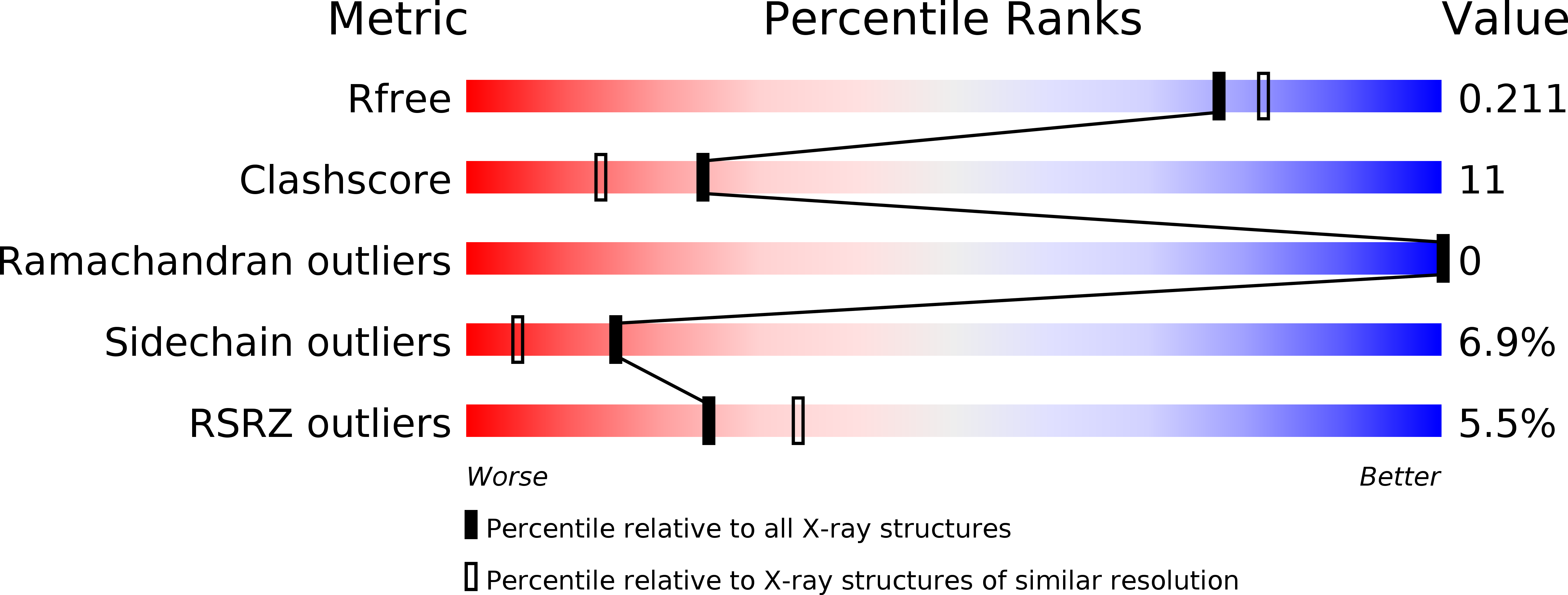
Resolution:
1.96 Å
R-Value Free:
0.21
R-Value Work:
0.17
R-Value Observed:
0.17
Space Group:
P 1
Macromolecular Entities
Polymer Type:polypeptide(L)
Description:Iron-dependent repressor ideR
Chain IDs:A, B
Chain Length:157
Number of Molecules:2
Biological Source:Mycobacterium tuberculosis
Modified Residue
Compound ID Chain ID Parent Comp ID Details 2D Image
CSO A CYS S-HYDROXYCYSTEINE
Primary Citation
Crystal structures, metal activation, and DNA-binding properties of two-domain IdeR from Mycobacterium tuberculosis
Biochemistry 46 436 447 (2007)
PMID: 17209554 DOI: 10.1021/bi0609826

Abstact

The iron-dependent regulator IdeR is a key transcriptional regulator of iron uptake in Mycobacterium tuberculosis. In order to increase our insight into the role of the SH3-like third domain of this essential regulator, the metal-binding and DNA-binding properties of two-domain IdeR (2D-IdeR) whose SH3-like domain has been truncated were characterized. The equilibrium dissociation constants for Co2+ and Ni2+ activation of 2D-IdeR for binding to the fxbA operator and the DNA-binding affinities of 2D-IdeR in the presence of excess metal ions were estimated using fluorescence spectroscopy. 2D-IdeR binds to fxbA operator DNA with similar affinity as full-length IdeR in the presence of excess metal ion. However, the Ni2+ concentrations required to activate 2D-IdeR for DNA binding appear to be smaller than that for full-length IdeR while the concentration of Co2+ required for activation remains the same. We have determined the crystal structures of Ni2+-activated 2D-IdeR at 1.96 A resolution and its double dimer complex with the mbtA-mbtB operator DNA in two crystal forms at 2.4 A and 2.6 A, the highest resolutions for DNA complexes for any structures of iron-dependent regulator family members so far. The 2D-IdeR-DNA complex structures confirm the specificity of Ser37 and Pro39 for thymine bases and suggest preferential contacts of Gln43 to cytosine bases of the DNA. In addition, our 2D-IdeR structures reveal a remarkable property of the TEV cleavage sequence remaining after removal of the C-terminal His6. This C-terminal tail promotes crystal contacts by forming a beta-sheet with the corresponding tail of neighboring subunits in two unrelated structures of 2D-IdeR, one with and one without DNA. The contact-promoting properties of this C-terminal TEV cleavage sequence may be beneficial for crystallizing other proteins.

Legend

Protein

Chemical

Disease

Primary Citation of related structures