2ISS image
Deposition Date 2006-10-18
Release Date 2007-01-02
Last Version Date 2024-10-16
Entry Detail
PDB ID:
2ISS
Title:
Structure of the PLP synthase Holoenzyme from Thermotoga maritima
Biological Source:
Source Organism:
Host Organism:
Method Details:
Experimental Method:
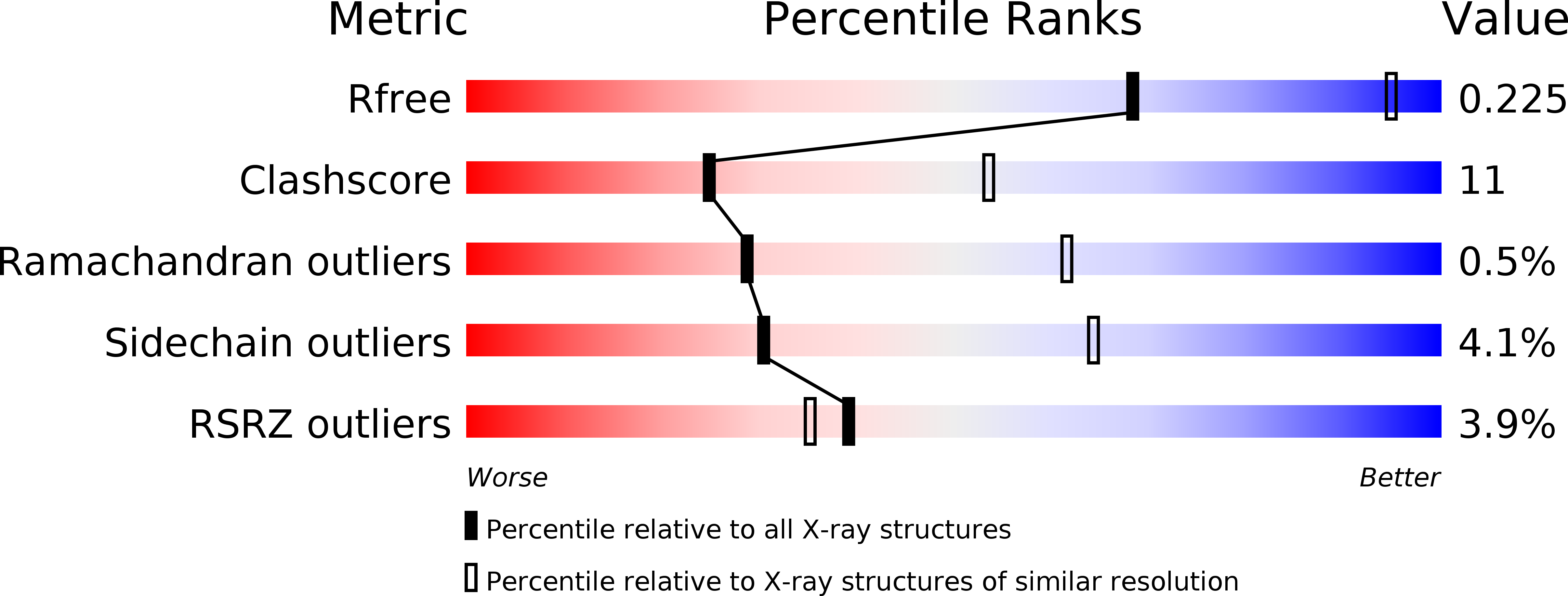
Resolution:
2.90 Å
R-Value Free:
0.23
R-Value Work:
0.21
R-Value Observed:
0.21
Space Group:
I 2 2 2
Macromolecular Entities
Structures with similar UniProt ID
Protein Blast
Polymer Type:polypeptide(L)
Molecule:Pyridoxal biosynthesis lyase pdxS
Gene (Uniprot):pdxS
Chain IDs:A, B, C
Chain Length:313
Number of Molecules:3
Biological Source:Thermotoga maritima
Structures with similar UniProt ID
Protein Blast
Polymer Type:polypeptide(L)
Molecule:Glutamine amidotransferase subunit pdxT
Gene (Uniprot):pdxT
Chain IDs:D, E, F
Chain Length:208
Number of Molecules:3
Biological Source:Thermotoga maritima
Primary Citation
Structural Insights into the Mechanism of the PLP Synthase Holoenzyme from Thermotoga maritima
Biochemistry 45 14609 14620 (2006)
PMID: 17144654 DOI: 10.1021/bi061464y

Abstact

Pyridoxal 5'-phosphate (PLP) is the biologically active form of vitamin B6 and is an important cofactor for several of the enzymes involved in the metabolism of amine-containing natural products such as amino acids and amino sugars. The PLP synthase holoenzyme consists of two subunits: YaaD catalyzes the condensation of ribulose 5-phosphate, glyceraldehyde-3-phosphate, and ammonia, and YaaE catalyzes the production of ammonia from glutamine. Here we describe the structure of the PLP synthase complex (YaaD-YaaE) from Thermotoga maritima at 2.9 A resolution. This complex consists of a core of 12 YaaD monomers with 12 noninteracting YaaE monomers attached to the core. Compared with the previously published structure of PdxS (a YaaD ortholog in Geobacillus stearothermophilus), the N-terminus (1-18), which includes helix alpha0, the beta2-alpha2 loop (46-56), which includes new helix alpha2a, and the C-terminus (270-280) of YaaD are ordered in the complex but disordered in PdxS. A ribulose 5-phosphate is bound to YaaD via an imine with Lys82. Previous studies have demonstrated a similar imine at Lys149 and not at Lys81 (equivalent to Lys150 and Lys82 in T. maritima) for the Bacillus subtilis enzyme suggesting the possibility that two separate sites on YaaD are involved in PLP formation. A phosphate from the crystallization solution is found bound to YaaD and also serves as a marker for a possible second active site. An ammonia channel that connects the active site of YaaE with the ribulose 5-phosphate binding site was identified. This channel is similar to one found in imidazole glycerol phosphate synthase; however, when the beta-barrels of the two complexes are superimposed, the glutaminase domains are rotated by about 180 degrees with respect to each other.

Legend

Protein

Chemical

Disease

Primary Citation of related structures
Feedback Form
Name
Email
Institute
Feedback