2IRV image
Deposition Date 2006-10-16
Release Date 2006-10-31
Last Version Date 2024-02-21
Entry Detail
PDB ID:
2IRV
Title:
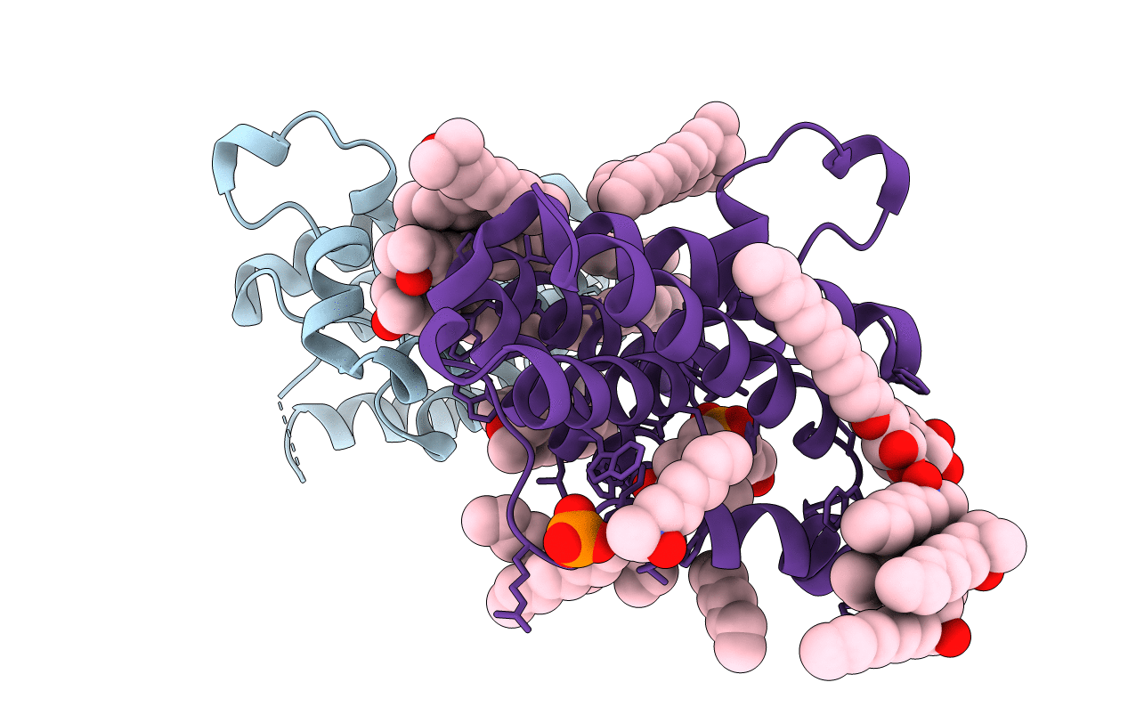
Crystal structure of GlpG, a rhomboid intramembrane serine protease
Biological Source:
Source Organism:
Host Organism:
Method Details:
Experimental Method:
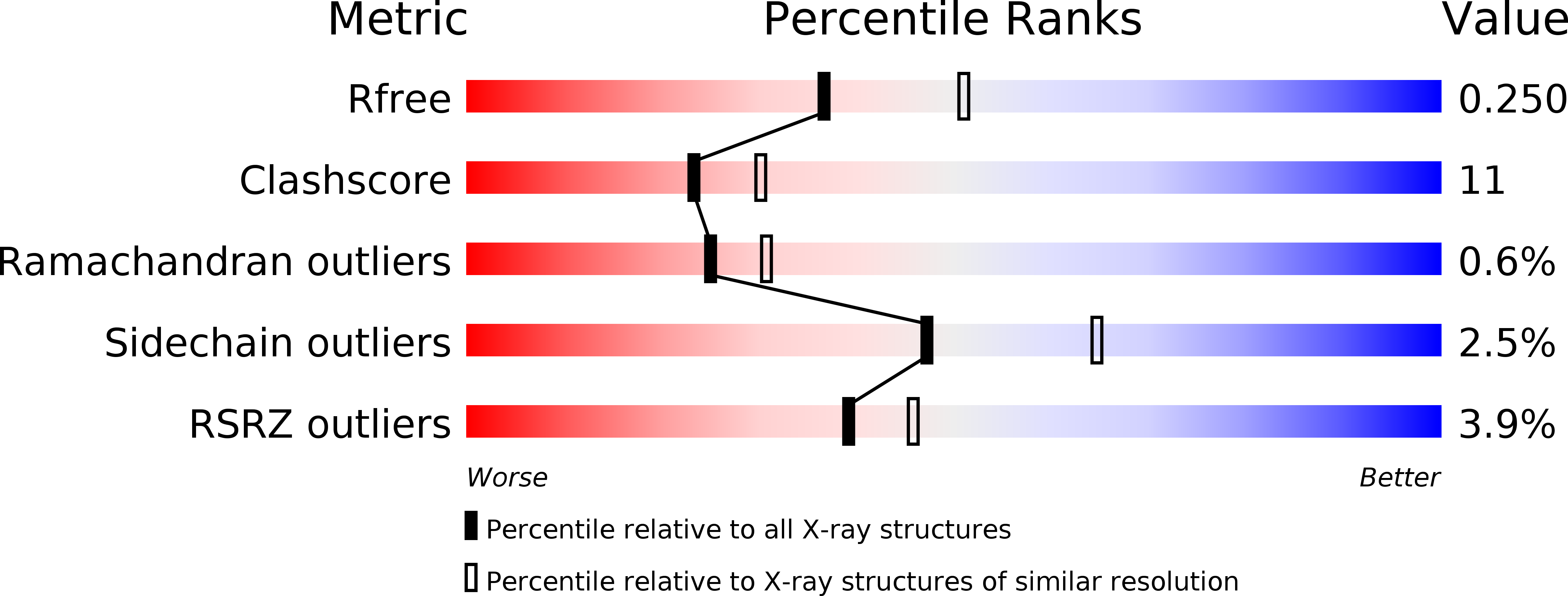
Resolution:
2.30 Å
R-Value Free:
0.26
R-Value Work:
0.22
Space Group:
P 1 21 1
Macromolecular Entities
Structures with similar UniProt ID
Protein Blast
Polymer Type:polypeptide(L)
Molecule:Protein glpG
Gene (Uniprot):glpG
Chain IDs:A, B
Chain Length:182
Number of Molecules:2
Biological Source:Escherichia coli
Primary Citation
Structural basis for intramembrane proteolysis by rhomboid serine proteases.
Proc.Natl.Acad.Sci.Usa 104 462 466 (2007)
PMID: 17190827 DOI: 10.1073/pnas.0609773104

Abstact

Intramembrane proteases catalyze peptide bond cleavage of integral membrane protein substrates. This activity is crucial for many biological and pathological processes. Rhomboids are evolutionarily widespread intramembrane serine proteases. Here, we present the 2.3-A-resolution crystal structure of a rhomboid from Escherichia coli. The enzyme has six transmembrane helices, five of which surround a short TM4, which starts deep within the membrane at the catalytic serine residue. Thus, the catalytic serine is in an externally exposed cavity, which provides a hydrophilic environment for proteolysis. Our results reveal a mechanism to enable water-dependent catalysis at the depth of the hydrophobic milieu of the membrane and suggest how substrates gain access to the sequestered rhomboid active site.

Legend

Protein

Chemical

Disease

Primary Citation of related structures