2IP4 image
Deposition Date 2006-10-11
Release Date 2007-10-30
Last Version Date 2024-11-13
Entry Detail
PDB ID:
2IP4
Keywords:
Title:
Crystal Structure of Glycinamide Ribonucleotide Synthetase from Thermus thermophilus HB8
Biological Source:
Source Organism:
Host Organism:
Method Details:
Experimental Method:
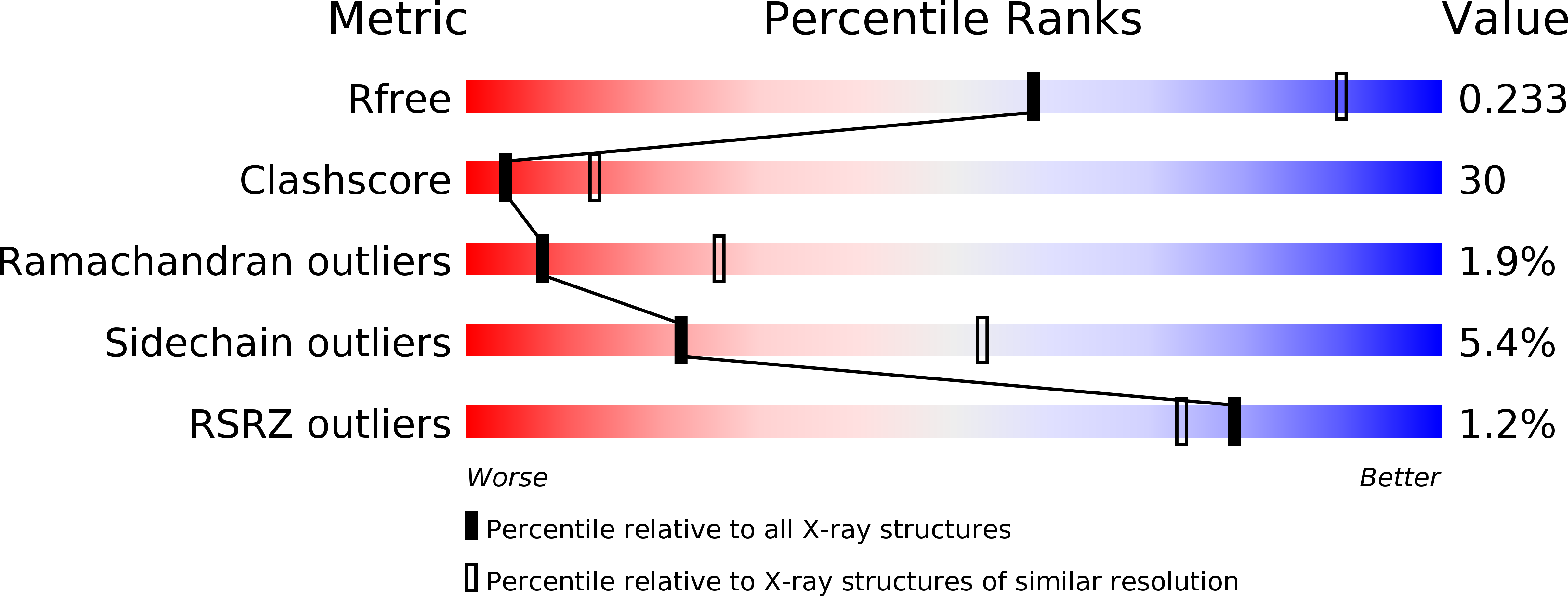
Resolution:
2.80 Å
R-Value Free:
0.23
R-Value Work:
0.21
R-Value Observed:
0.22
Space Group:
P 21 21 21
Macromolecular Entities
Polymer Type:polypeptide(L)
Molecule:Phosphoribosylamine--glycine ligase
Gene (Uniprot):purD
Chain IDs:A, B
Chain Length:417
Number of Molecules:2
Biological Source:Thermus thermophilus
Modified Residue
Compound ID Chain ID Parent Comp ID Details 2D Image
MSE A MET SELENOMETHIONINE
Ligand Molecules
Primary Citation
Crystal structures of glycinamide ribonucleotide synthetase, PurD, from thermophilic eubacteria
J.Biochem. 148 429 438 (2010)
PMID: 20716513 DOI: 10.1093/jb/mvq088

Abstact

Glycinamide ribonucleotide synthetase (GAR-syn, PurD) catalyses the second reaction of the purine biosynthetic pathway; the conversion of phosphoribosylamine, glycine and ATP to glycinamide ribonucleotide (GAR), ADP and Pi. In the present study, crystal structures of GAR-syn's from Thermus thermophilus, Geobacillus kaustophilus and Aquifex aeolicus were determined in apo forms. Crystal structures in ligand-bound forms were also determined for G. kaustophilus and A. aeolicus proteins. In general, overall structures of GAR-syn's are similar to each other. However, the orientations of the B domains are varied among GAR-syn's and the MD simulation suggested the mobility of the B domain. Furthermore, it was demonstrated that the B loop in the B domain fixes the position of the β- and γ- phosphate groups of the bound ATP. The structures of GAR-syn's and the bound ligands were compared with each other in detail, and structures of GAR-syn's with full ligands, as well as the possible reaction mechanism, were proposed.

Legend

Protein

Chemical

Disease

Primary Citation of related structures