2IOR image
Deposition Date 2006-10-10
Release Date 2006-11-21
Last Version Date 2023-08-30
Entry Detail
PDB ID:
2IOR
Keywords:
Title:
Crystal Structure of the N-terminal Domain of HtpG, the Escherichia coli Hsp90, Bound to ADP
Biological Source:
Source Organism:
Escherichia coli (Taxon ID: 562)
Method Details:
Experimental Method:
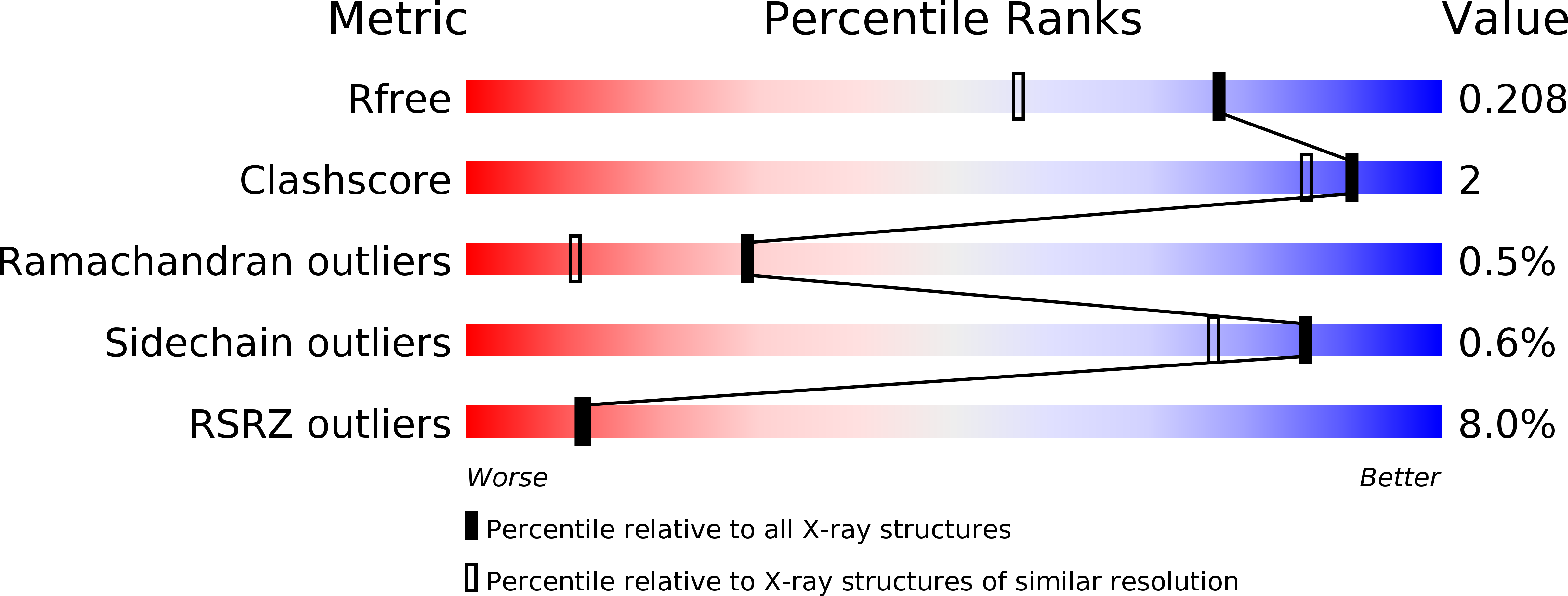
Resolution:
1.65 Å
R-Value Free:
0.19
R-Value Work:
0.16
R-Value Observed:
0.16
Space Group:
P 21 21 21
Macromolecular Entities
Structures with similar UniProt ID
Protein Blast
Polymer Type:polypeptide(L)
Molecule:Chaperone protein htpG
Gene (Uniprot):htpG
Chain IDs:A
Chain Length:235
Number of Molecules:1
Biological Source:Escherichia coli
Primary Citation
Structural Analysis of E. coli hsp90 reveals dramatic nucleotide-dependent conformational rearrangements.
Cell(Cambridge,Mass.) 127 329 340 (2006)
PMID: 17055434 DOI: 10.1016/j.cell.2006.09.027

Abstact

In eukaryotes, the ubiquitous and abundant members of the 90 kilodalton heat-shock protein (hsp90) chaperone family facilitate the folding and conformational changes of a broad array of proteins important in cell signaling, proliferation, and survival. Here we describe the effects of nucleotides on the structure of full-length HtpG, the Escherichia coli hsp90 ortholog. By electron microscopy, the nucleotide-free, AMPPNP bound, and ADP bound states of HtpG adopt completely distinct conformations. Structural characterization of nucleotide-free and ADP bound HtpG was extended to higher resolution by X-ray crystallography. In the absence of nucleotide, HtpG exhibits an "open" conformation in which the three domains of each monomer present hydrophobic elements into the large cleft formed by the dimer. By contrast, ADP binding drives dramatic conformational changes that allow these hydrophobic elements to converge and shield each other from solvent, suggesting a mechanism by which nucleotides could control client protein binding and release.

Legend

Protein

Chemical

Disease

Primary Citation of related structures