2INN image
Deposition Date 2006-10-08
Release Date 2007-01-16
Last Version Date 2024-11-13
Entry Detail
PDB ID:
2INN
Keywords:
Title:
Structure of the Phenol Hydroxyalse-Regulatory Protein Complex
Biological Source:
Source Organism:
Host Organism:
Method Details:
Experimental Method:
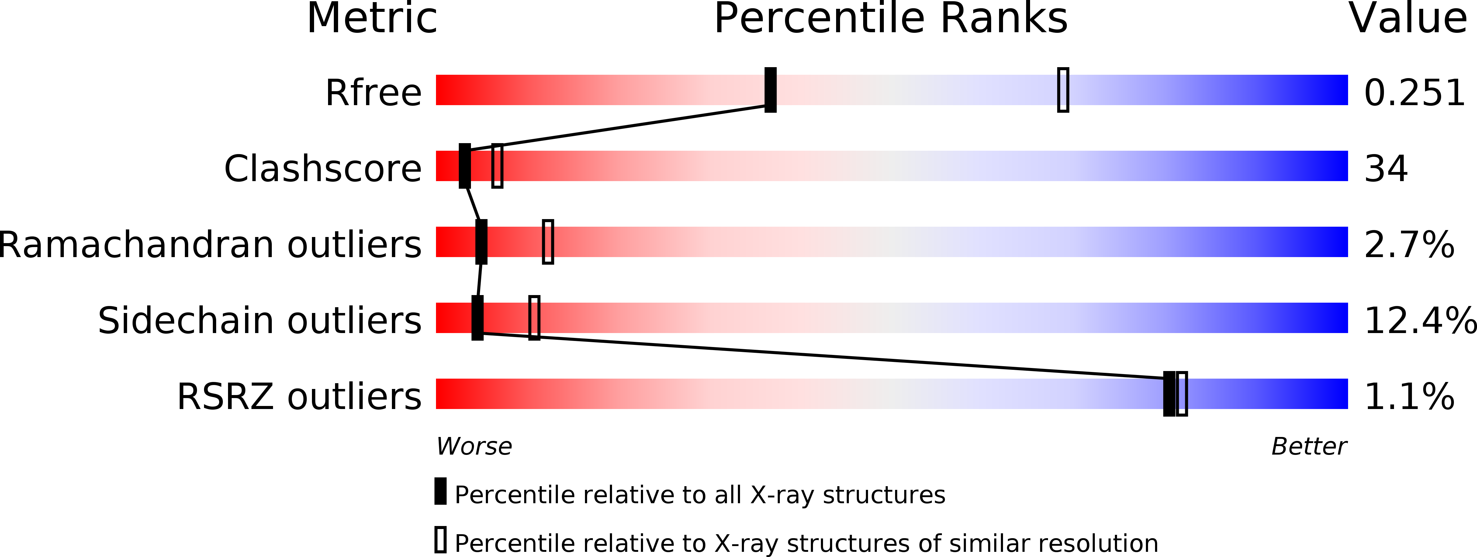
Resolution:
2.70 Å
R-Value Free:
0.25
R-Value Work:
0.20
R-Value Observed:
0.20
Space Group:
P 21 21 21
Macromolecular Entities
Structures with similar UniProt ID
Protein Blast
Polymer Type:polypeptide(L)
Molecule:Phenol hydroxylase component phN
Gene (Uniprot):phN
Chain IDs:A, B
Chain Length:511
Number of Molecules:2
Biological Source:Pseudomonas stutzeri
Structures with similar UniProt ID
Protein Blast
Polymer Type:polypeptide(L)
Molecule:Phenol hydroxylase component phL
Gene (Uniprot):phL
Chain IDs:C, D
Chain Length:333
Number of Molecules:2
Biological Source:Pseudomonas stutzeri
Structures with similar UniProt ID
Protein Blast
Polymer Type:polypeptide(L)
Molecule:Phenol hydroxylase component phO
Gene (Uniprot):phO
Chain IDs:E, F
Chain Length:119
Number of Molecules:2
Biological Source:Pseudomonas stutzeri
Structures with similar UniProt ID
Protein Blast
Polymer Type:polypeptide(L)
Molecule:Phenol hydroxylase component phM
Gene (Uniprot):phM
Chain IDs:G (auth: L)
Chain Length:89
Number of Molecules:1
Biological Source:Pseudomonas stutzeri
Modified Residue
Compound ID Chain ID Parent Comp ID Details 2D Image
MSE A MET SELENOMETHIONINE
Primary Citation
X-ray Structure of a Hydroxylase-Regulatory Protein Complex from a Hydrocarbon-Oxidizing Multicomponent Monooxygenase, Pseudomonas sp. OX1 Phenol Hydroxylase.
Biochemistry 45 15392 15404 (2006)
PMID: 17176061 DOI: 10.1021/bi0618969

Abstact

Phenol hydroxylase (PH) belongs to a family of bacterial multicomponent monooxygenases (BMMs) with carboxylate-bridged diiron active sites. Included are toluene/o-xylene (ToMO) and soluble methane (sMMO) monooxygenase. PH hydroxylates aromatic compounds, but unlike sMMO, it cannot oxidize alkanes despite having a similar dinuclear iron active site. Important for activity is formation of a complex between the hydroxylase and a regulatory protein component. To address how structural features of BMM hydroxylases and their component complexes may facilitate the catalytic mechanism and choice of substrate, we determined X-ray structures of native and SeMet forms of the PH hydroxylase (PHH) in complex with its regulatory protein (PHM) to 2.3 A resolution. PHM binds in a canyon on one side of the (alphabetagamma)2 PHH dimer, contacting alpha-subunit helices A, E, and F approximately 12 A above the diiron core. The structure of the dinuclear iron center in PHH resembles that of mixed-valent MMOH, suggesting an Fe(II)Fe(III) oxidation state. Helix E, which comprises part of the iron-coordinating four-helix bundle, has more pi-helical character than analogous E helices in MMOH and ToMOH lacking a bound regulatory protein. Consequently, conserved active site Thr and Asn residues translocate to the protein surface, and an approximately 6 A pore opens through the four-helix bundle. Of likely functional significance is a specific hydrogen bond formed between this Asn residue and a conserved Ser side chain on PHM. The PHM protein covers a putative docking site on PHH for the PH reductase, which transfers electrons to the PHH diiron center prior to O2 activation, suggesting that the regulatory component may function to block undesired reduction of oxygenated intermediates during the catalytic cycle. A series of hydrophobic cavities through the PHH alpha-subunit, analogous to those in MMOH, may facilitate movement of the substrate to and/or product from the active site pocket. Comparisons between the ToMOH and PHH structures provide insights into their substrate regiospecificities.

Legend

Protein

Chemical

Disease

Primary Citation of related structures