2ILN image
Deposition Date 2006-10-03
Release Date 2007-04-10
Last Version Date 2024-11-20
Entry Detail
PDB ID:
2ILN
Title:
Crystal structure of the Bowman-Birk inhibitor from snail medic seeds in complex with bovine trypsin
Biological Source:
Source Organism:
Bos taurus (Taxon ID: 9913)
Medicago scutellata (Taxon ID: 36901)
Method Details:
Experimental Method:
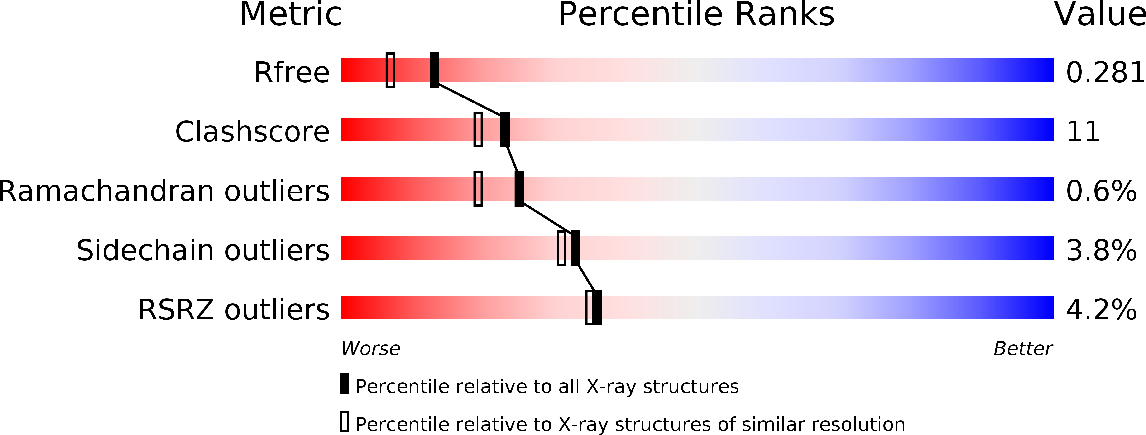
Resolution:
2.00 Å
R-Value Free:
0.28
R-Value Work:
0.23
R-Value Observed:
0.23
Space Group:
P 41
Macromolecular Entities
Structures with similar UniProt ID
Protein Blast
Polymer Type:polypeptide(L)
Molecule:Cationic trypsin
Gene (Uniprot):PRSS1
Chain IDs:A, B
Chain Length:223
Number of Molecules:2
Biological Source:Bos taurus
Structures with similar UniProt ID
Protein Blast
Polymer Type:polypeptide(L)
Molecule:Bowman-Birk type proteinase inhibitor
Chain IDs:C (auth: I)
Chain Length:62
Number of Molecules:1
Biological Source:Medicago scutellata
Primary Citation
Crystal structure of the anticarcinogenic Bowman-Birk inhibitor from snail medic (Medicago scutellata) seeds complexed with bovine trypsin
J.Struct.Biol. 158 71 79 (2007)
PMID: 17142058 DOI: 10.1016/j.jsb.2006.10.017

Abstact

The structure of the ternary complex of the anticarcinogenic Bowman-Birk protease inhibitor purified from snail medic (Medicago scutellata) seeds (MSTI) and two molecules of bovine trypsin has been solved by X-ray diffraction analysis of single crystals to a resolution of 2.0 A. This is the highest resolution model of a ternary complex of this type currently available. The two binding loops of the MSTI differ in only one amino acid and have in both cases an arginine in position P1. The distances between the residues of the inhibitor at the binding interface and the trypsin side chains that recognize them are almost identical in the two sites. When compared to the NMR model of the uncomplexed MSTI, the inhibitor in the functional assembly with trypsin shows the largest differences in the two P2' residues. Compared with the similar ternary complex of the soybean trypsin inhibitor, this model shows very small differences in the polypeptide chain of the trypsin binding sites and its largest difference in the area between Asp 26 and His 32 of the MSTI which in the soybean inhibitor has an extra Leu inserted in position 29.

Legend

Protein

Chemical

Disease

Primary Citation of related structures
Feedback Form
Name
Email
Institute
Feedback