Deposition Date
2006-10-02
Release Date
2006-11-21
Last Version Date
2023-08-30
Entry Detail
PDB ID:
2IK8
Keywords:
Title:
Crystal structure of the heterodimeric complex of human RGS16 and activated Gi alpha 1
Biological Source:
Source Organism(s):
Homo sapiens (Taxon ID: 9606)
Expression System(s):
Method Details:
Experimental Method:
Resolution:
2.71 Å
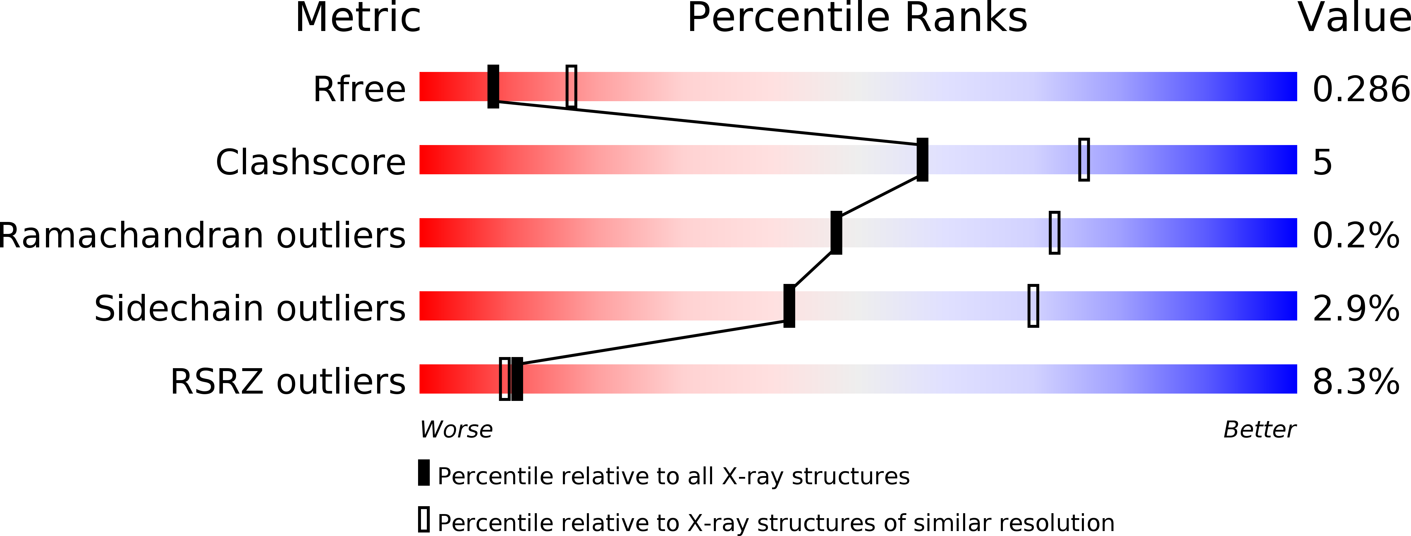
R-Value Free:
0.29
R-Value Work:
0.23
R-Value Observed:
0.23
Space Group:
P 21 21 21