Deposition Date
2006-09-27
Release Date
2006-10-31
Last Version Date
2024-10-30
Entry Detail
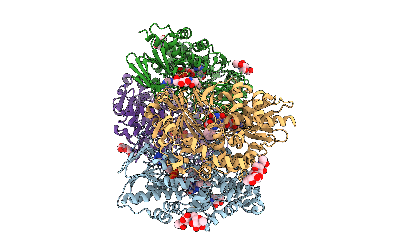
PDB ID:
2IID
Keywords:
Title:
Structure of L-amino acid oxidase from Calloselasma rhodostoma in complex with L-phenylalanine
Biological Source:
Source Organism(s):
Calloselasma rhodostoma (Taxon ID: 8717)
Method Details:
Experimental Method:
Resolution:
1.80 Å
R-Value Free:
0.21
R-Value Work:
0.17
R-Value Observed:
0.17
Space Group:
P 1 21 1