Deposition Date
2006-09-26
Release Date
2007-08-21
Last Version Date
2023-08-30
Entry Detail
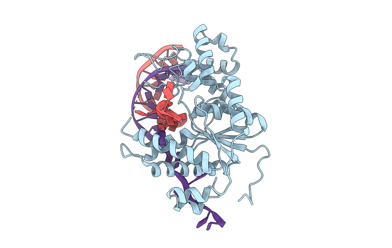
PDB ID:
2IHN
Keywords:
Title:
Co-crystal of Bacteriophage T4 RNase H with a fork DNA substrate
Biological Source:
Source Organism(s):
Enterobacteria phage T4 (Taxon ID: 10665)
Expression System(s):
Method Details:
Experimental Method:
Resolution:
3.00 Å
R-Value Free:
0.28
R-Value Work:
0.22
R-Value Observed:
0.22
Space Group:
P 21 21 21