Deposition Date
2006-09-26
Release Date
2007-01-02
Last Version Date
2023-08-30
Entry Detail
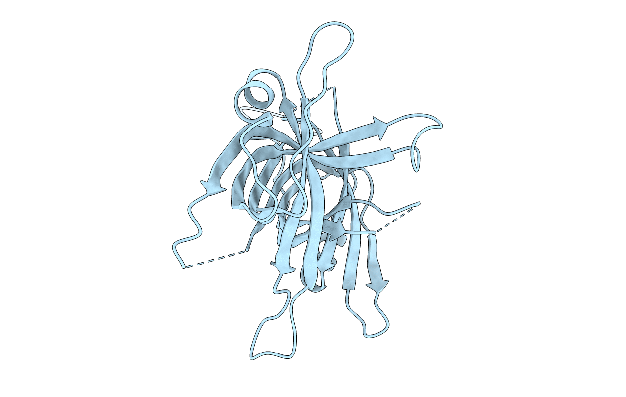
PDB ID:
2IHE
Keywords:
Title:
Crystal structure of wild-type single-stranded DNA binding protein from Thermus aquaticus
Biological Source:
Source Organism(s):
Thermus aquaticus (Taxon ID: 271)
Expression System(s):
Method Details:
Experimental Method:
Resolution:
2.10 Å
R-Value Free:
0.25
R-Value Work:
0.20
R-Value Observed:
0.20
Space Group:
C 1 2 1