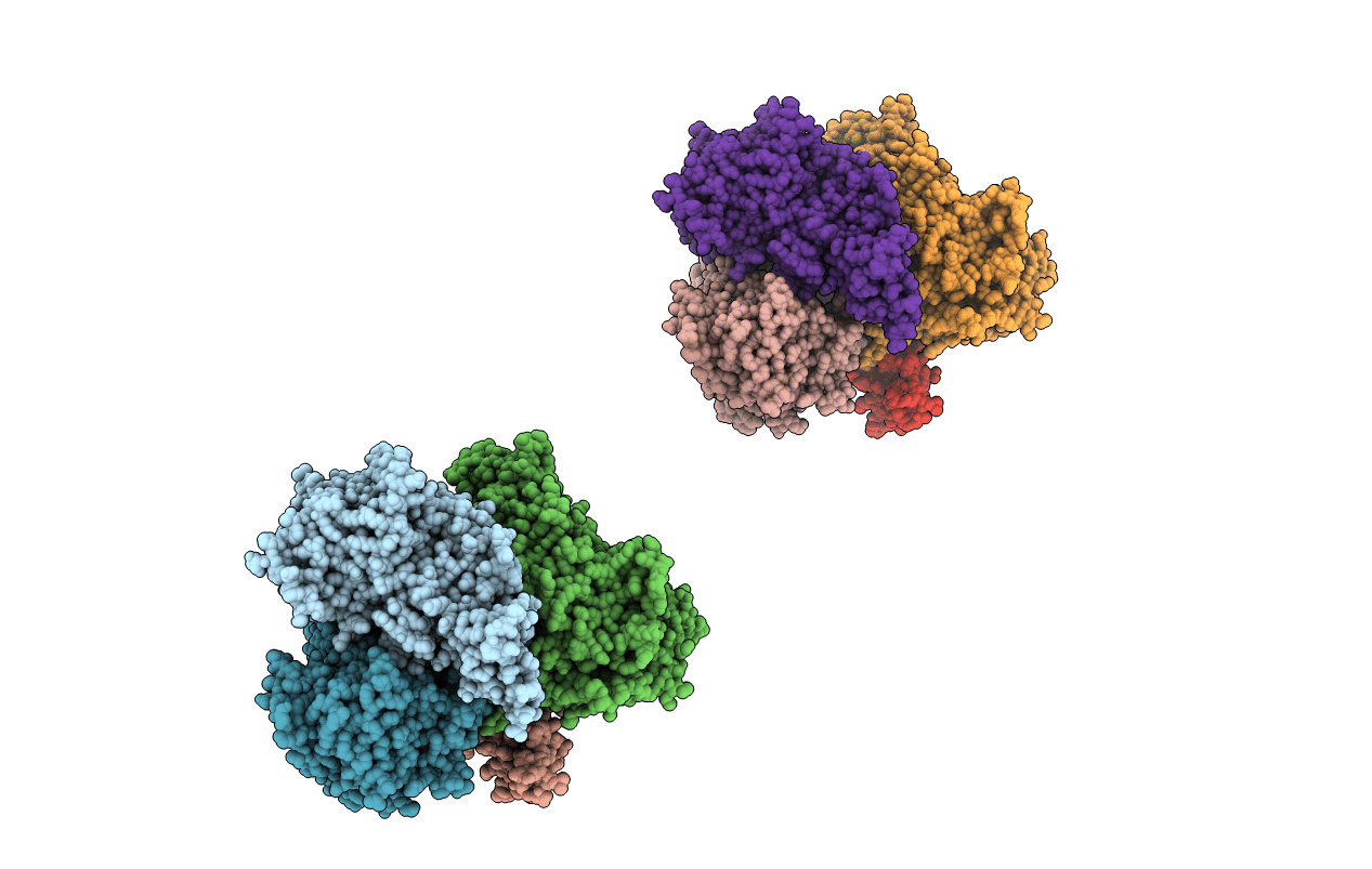
2IGO image
Entry Detail
PDB ID:
2IGO
Keywords:
Title:
Crystal structure of pyranose 2-oxidase H167A mutant with 2-fluoro-2-deoxy-D-glucose
Biological Source:
Source Organism:
PDB Version:
Deposition Date:
2006-09-22
Release Date:
2006-10-10
Method Details:
Experimental Method:
Resolution:
1.95 Å
R-Value Free:
0.22
R-Value Work:
0.18
R-Value Observed:
0.18
Space Group:
P 1 21 1
Macromolecular Entities
Polymer Type:polypeptide(L)
Description:Pyranose oxidase
Mutations:H167A
Chain IDs:A, B, C (auth: D), D (auth: C), E, F, G (auth: H), H (auth: G)
Chain Length:623
Number of Molecules:8
Biological Source:Trametes ochracea
Primary Citation
Structural basis for substrate binding and regioselective oxidation of monosaccharides at c3 by pyranose 2-oxidase.
J.Biol.Chem. 281 35104 35115 (2006)
PMID: 16984920 DOI: 10.1074/jbc.M604718200

Abstact

Pyranose 2-oxidase (P2Ox) participates in fungal lignin degradation by producing the H2O2 needed for lignin-degrading peroxidases. The enzyme oxidizes cellulose- and hemicellulose-derived aldopyranoses at C2 preferentially, but also on C3, to the corresponding ketoaldoses. To investigate the structural determinants of catalysis, covalent flavinylation, substrate binding, and regioselectivity, wild-type and mutant P2Ox enzymes were produced and characterized biochemically and structurally. Removal of the histidyl-FAD linkage resulted in a catalytically competent enzyme containing tightly, but noncovalently bound FAD. This mutant (H167A) is characterized by a 5-fold lower kcat, and a 35-mV lower redox potential, although no significant structural changes were seen in its crystal structure. In previous structures of P2Ox, the substrate loop (residues 452-457) covering the active site has been either disordered or in a conformation incompatible with carbohydrate binding. We present here the crystal structure of H167A in complex with a slow substrate, 2-fluoro-2-deoxy-D-glucose. Based on the details of 2-fluoro-2-deoxy-D-glucose binding in position for oxidation at C3, we also outline a probable binding mode for D-glucose positioned for regioselective oxidation at C2. The tentative determinant for discriminating between the two binding modes is the position of the O6 hydroxyl group, which in the C2-oxidation mode can make favorable interactions with Asp452 in the substrate loop and, possibly, a nearby arginine residue (Arg472). We also substantiate our hypothesis with steady-state kinetics data for the alanine replacements of Asp452 and Arg472 as well as the double alanine 452/472 mutant.

Legend

Protein

Chemical

Disease

Primary Citation of related structures