
Deposition Date
2006-09-21
Release Date
2006-10-03
Last Version Date
2023-08-30
Entry Detail
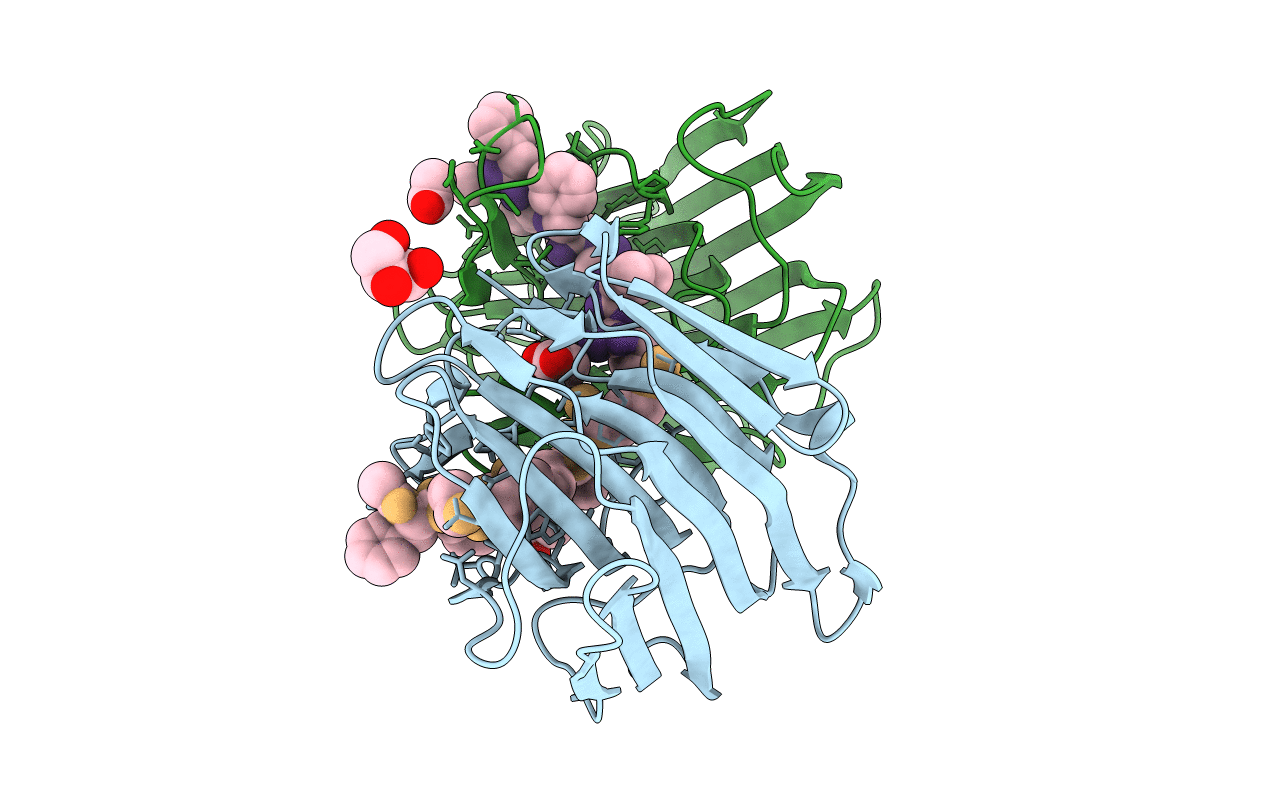
PDB ID:
2IFW
Keywords:
Title:
Crystal structure of scytalido-glutamic peptidase with a transition state analog inhibitor
Biological Source:
Source Organism(s):
Scytalidium lignicola (Taxon ID: 5539)
Expression System(s):
Method Details:
Experimental Method:
Resolution:
2.30 Å
R-Value Free:
0.26
R-Value Work:
0.22
R-Value Observed:
0.26
Space Group:
P 21 21 21
