2IES image
Deposition Date 2006-09-19
Release Date 2007-01-09
Last Version Date 2023-08-30
Entry Detail
PDB ID:
2IES
Keywords:
Title:
Crystal Structure of Aquifex aeolicus LpxC Complexed with Pyrophosphate
Biological Source:
Source Organism:
Host Organism:
Method Details:
Experimental Method:
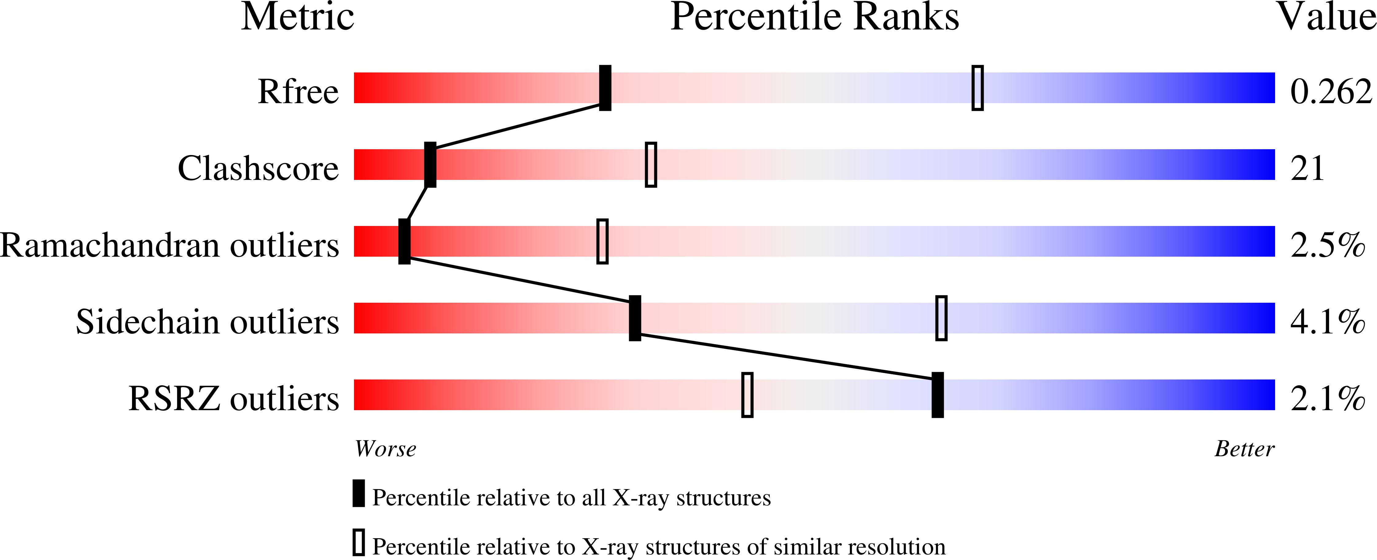
Resolution:
3.10 Å
R-Value Free:
0.27
R-Value Work:
0.23
Space Group:
P 61
Macromolecular Entities
Structures with similar UniProt ID
Protein Blast
Polymer Type:polypeptide(L)
Molecule:UDP-3-O-[3-hydroxymyristoyl] N-acetylglucosamine deacetylase
Gene (Uniprot):lpxC
Mutagens:C193A
Chain IDs:A, B
Chain Length:271
Number of Molecules:2
Biological Source:Aquifex aeolicus
Primary Citation
Binding of Uridine 5-Diphosphate in the Basic Patch of the Zinc Metalloenzyme Deacetylase LpxC and Implications for Substrate Binding
Biochemistry 45 15216 15223 (2006)
PMID: 17176043 DOI: 10.1021/bi0619021

Abstact

LpxC is a zinc metalloenzyme that catalyzes the first committed step in the biosynthesis of lipid A, a vital component of the outer membrane of Gram-negative bacteria. Accordingly, the inhibition of LpxC is an attractive strategy for the treatment of Gram-negative bacterial infections. Here, we report the 2.7 A resolution X-ray crystal structure of LpxC from Aquifex aeolicus complexed with uridine 5'-diphosphate (UDP), and the 3.1 A resolution structure of LpxC complexed with pyrophosphate. The X-ray crystal structure of the LpxC-UDP complex provides the first view of interactions likely to be exploited by the substrate UDP group in the "basic patch" of the active site. The diphosphate group of UDP makes hydrogen bond interactions with strictly conserved residue K239 as well as solvent molecules. The ribose moiety of UDP interacts with partially conserved residue E197. The UDP uracil group hydrogen bonds with both the backbone NH group and the backbone carbonyl group of E160, and with the backbone NH group of K162 through an intervening water molecule. Finally, the alpha-phosphate and uracil groups of UDP interact with R143 and R262 through intervening water molecules. The structure of LpxC complexed with pyrophosphate reveals generally similar intermolecular interactions in the basic patch. Unexpectedly, diphosphate binding in both complexes is accompanied by coordination to an additional zinc ion, resulting in the identification of a new metal-binding site termed the E-site. The structures of the LpxC-UDP and LpxC-pyrophosphate complexes provide new insights with regard to substrate recognition in the basic patch and metal ion coordination in the active site of LpxC.

Legend

Protein

Chemical

Disease

Primary Citation of related structures