Deposition Date
2006-09-19
Release Date
2006-12-26
Last Version Date
2024-11-20
Entry Detail
PDB ID:
2IEI
Keywords:
Title:
Crystal structure of rabbit muscle glycogen phosphorylase in complex with 3,4-dihydro-2-quinolone
Biological Source:
Source Organism(s):
Oryctolagus cuniculus (Taxon ID: 9986)
Method Details:
Experimental Method:
Resolution:
1.91 Å
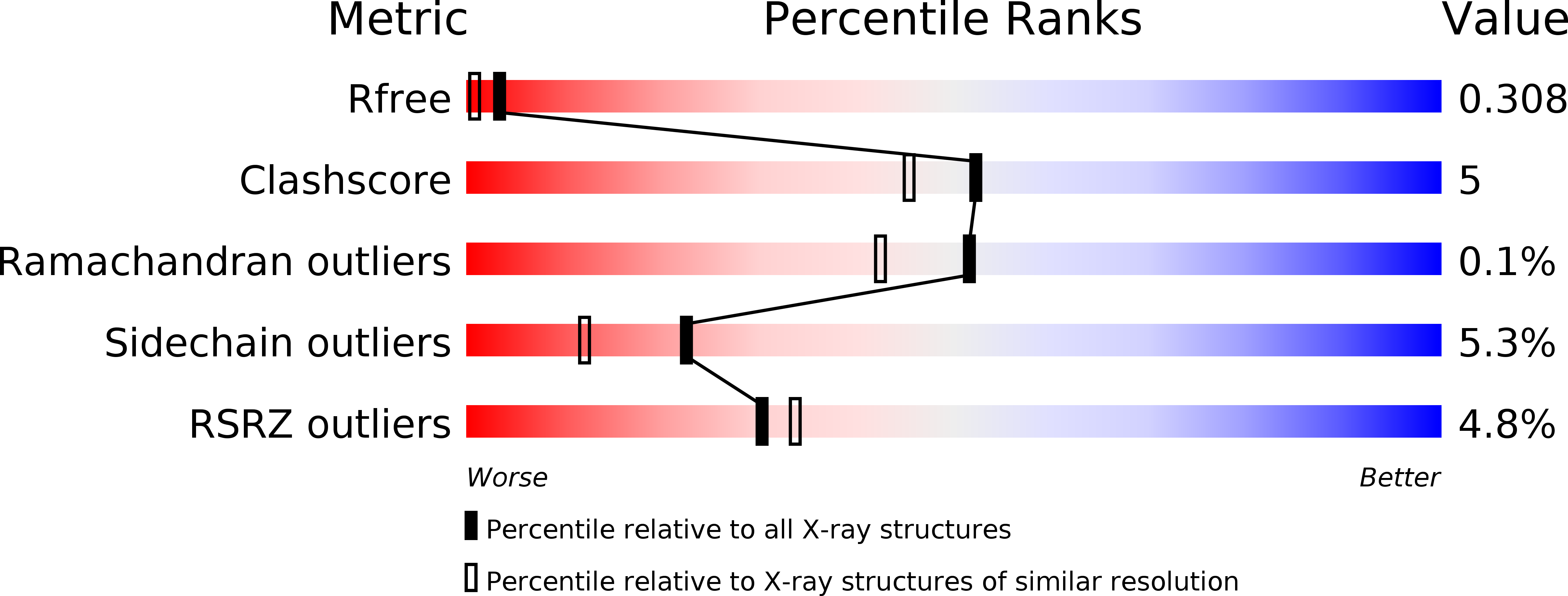
R-Value Free:
0.29
R-Value Work:
0.25
R-Value Observed:
0.25
Space Group:
P 21 21 21