Deposition Date
2006-09-15
Release Date
2006-11-14
Last Version Date
2023-08-30
Entry Detail
PDB ID:
2IDO
Keywords:
Title:
Structure of the E. coli Pol III epsilon-Hot proofreading complex
Biological Source:
Source Organism(s):
Escherichia coli (Taxon ID: 562)
Enterobacteria phage (Taxon ID: 10678)
Enterobacteria phage (Taxon ID: 10678)
Expression System(s):
Method Details:
Experimental Method:
Resolution:
2.10 Å
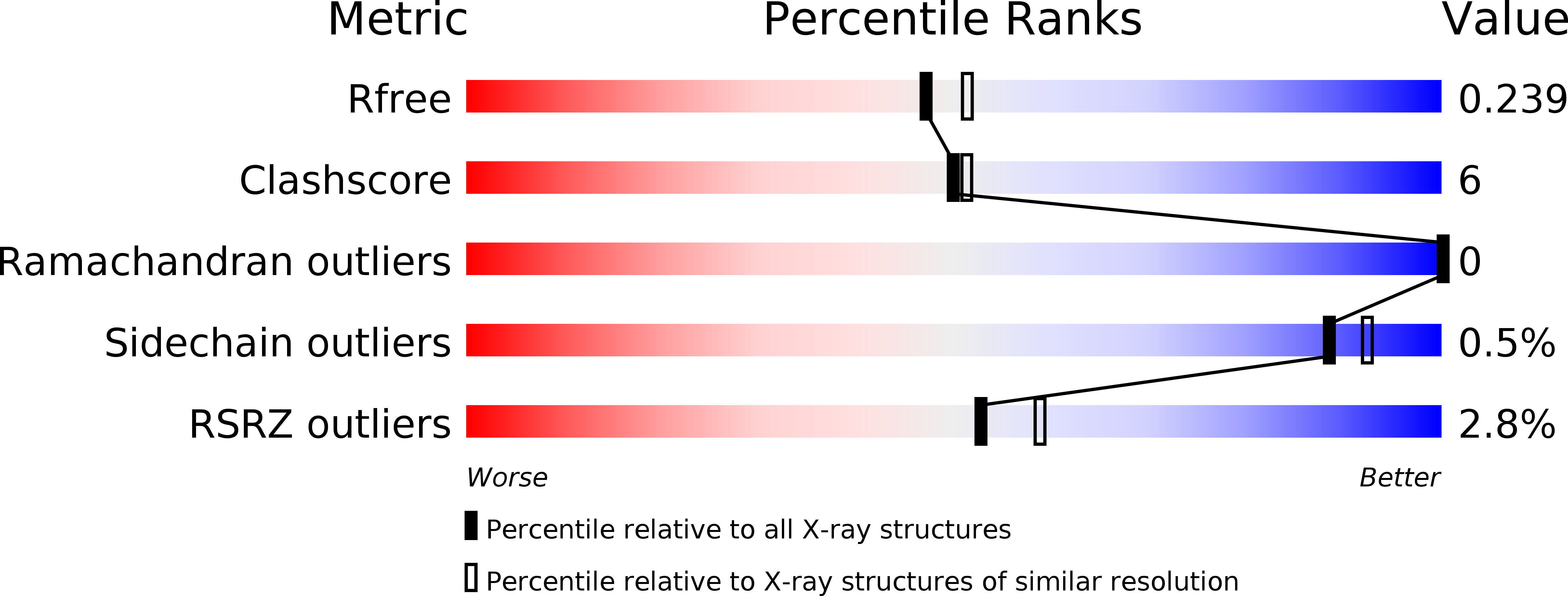
R-Value Free:
0.24
R-Value Work:
0.20
R-Value Observed:
0.20
Space Group:
P 43 21 2