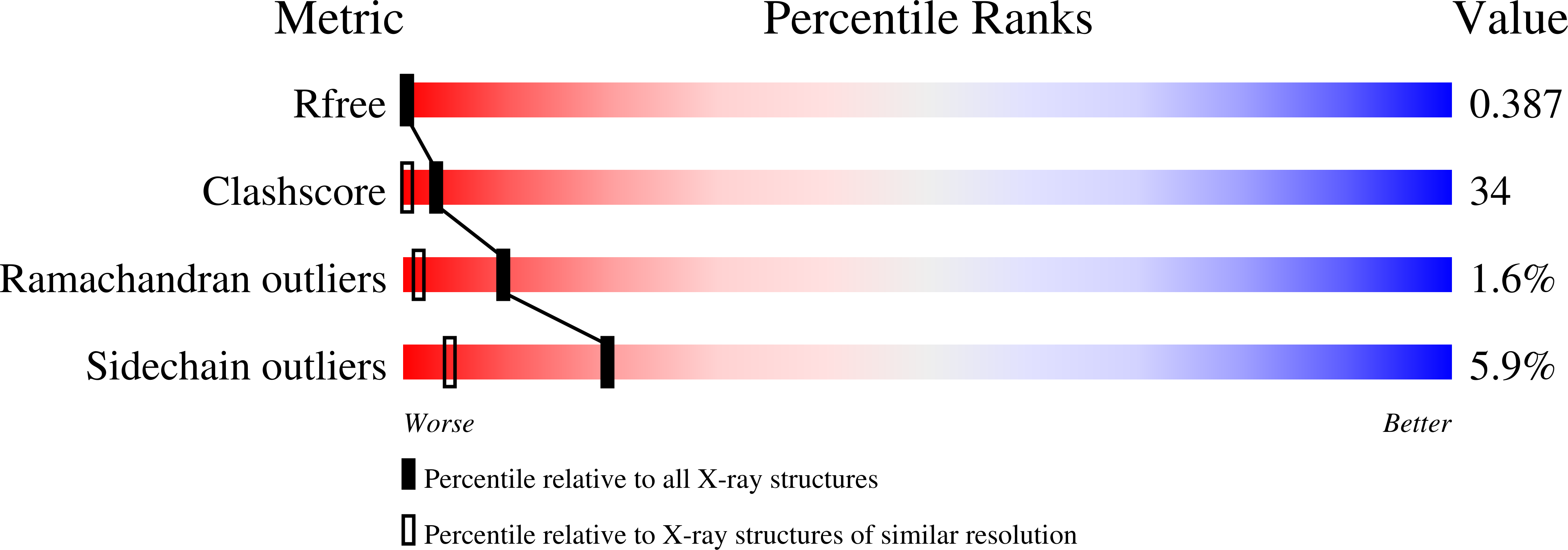
2ID9 image
Deposition Date 2006-09-14
Release Date 2007-09-25
Last Version Date 2023-08-30
Entry Detail
PDB ID:
2ID9
Title:
1.85 A Structure of T87I/Y106W Phosphono-CheY
Biological Source:
Source Organism:
Escherichia coli (Taxon ID: 562)
Host Organism:
Method Details:
Experimental Method:
Resolution:
1.75 Å
R-Value Free:
0.20
R-Value Work:
0.17
Space Group:
P 21 21 21
Macromolecular Entities
Polymer Type:polypeptide(L)
Molecule:Chemotaxis protein cheY
Gene (Uniprot):cheY
Mutations:D57(CyQ) T87I Y106W
Chain IDs:A
Chain Length:128
Number of Molecules:1
Biological Source:Escherichia coli
Modified Residue
Compound ID Chain ID Parent Comp ID Details 2D Image
CYQ A CYS ?
Primary Citation
The structures of T87I phosphono-CheY and T87I/Y106W phosphono-CheY help to explain their binding affinities to the FliM and CheZ peptides.
Arch.Biochem.Biophys. 479 105 113 (2008)
PMID: 18801331 DOI: 10.1016/j.abb.2008.08.019

Abstact

CheY is a response regulator in bacterial chemotaxis. Escherichia coli CheY mutants T87I and T87I/Y106W CheY are phosphorylatable on Asp57 but unable to generate clockwise rotation of the flagella. To understand this phenotype in terms of structure, stable analogs of the two CheY-P mutants were synthesized: T87I phosphono-CheY and T87I phosphono-CheY. Dissociation constants for peptides derived from flagellar motor protein FliM and phosphatase CheZ were determined for phosphono-CheY and the two mutants. The peptides bind phosphono-CheY almost as strongly as CheY-P; however, they do not bind T87I phosphono-CheY or T87I/Y106W phosphono-CheY, implying that the mutant proteins cannot bind FliM or CheZ tightly in vivo. The structures of T87I phosphono-CheY and T87I/Y106W phosphono-CheY were solved to resolutions of 1.8 and 2.4A, respectively. The increased bulk of I87 forces the side-chain of Y106 or W106, into a more solvent-accessible conformation, which occludes the peptide-binding site.

Legend

Protein

Chemical

Disease

Primary Citation of related structures