Deposition Date
2006-09-14
Release Date
2007-05-01
Last Version Date
2024-12-25
Entry Detail
PDB ID:
2ID4
Keywords:
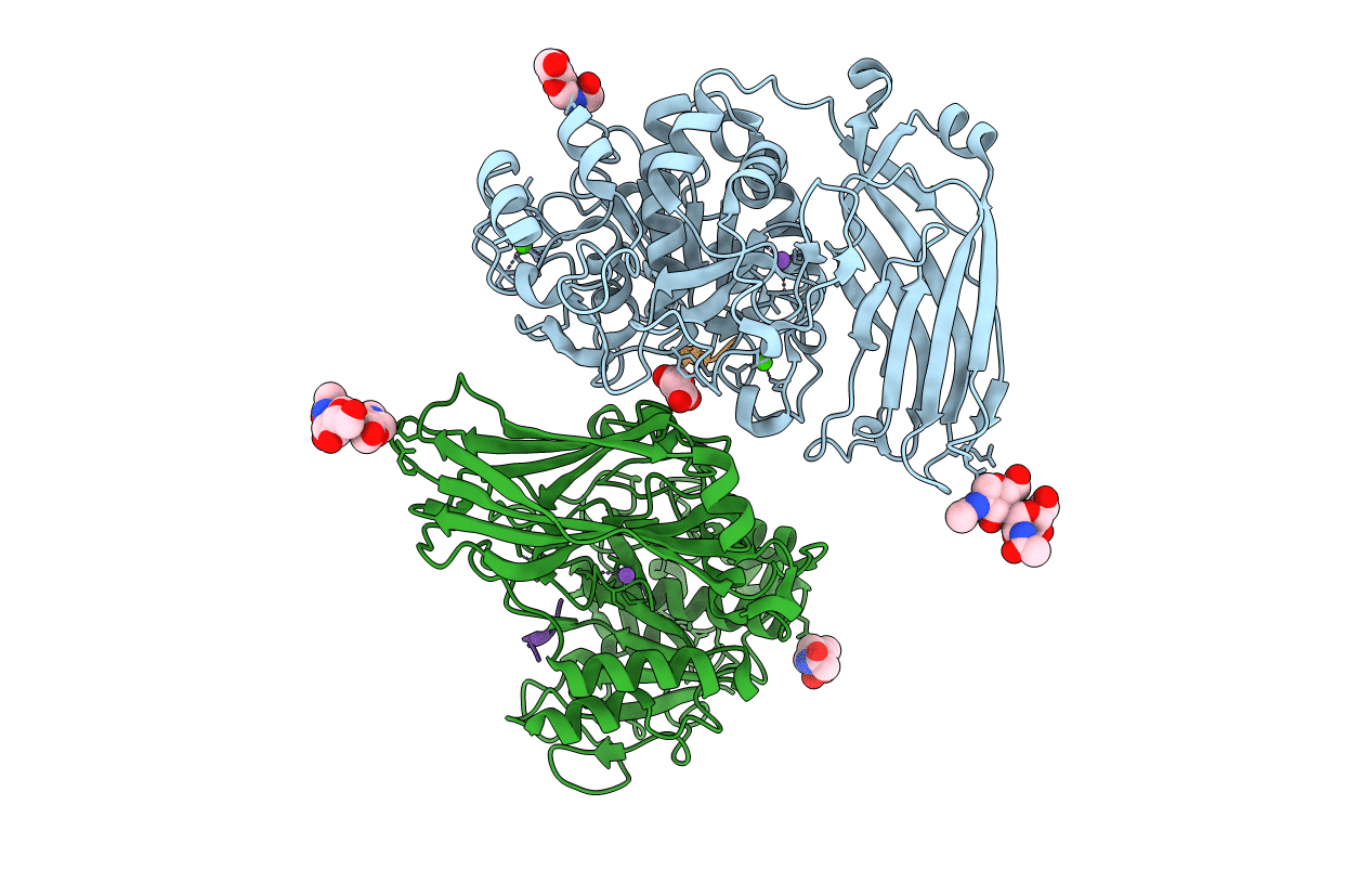
Title:
The 1.9 A structure of Kex2 in complex with an Ac-R-E-R-K-chloromethyl ketone inhibitor.
Biological Source:
Source Organism(s):
Saccharomyces cerevisiae (Taxon ID: 4932)
Expression System(s):
Method Details:
Experimental Method:
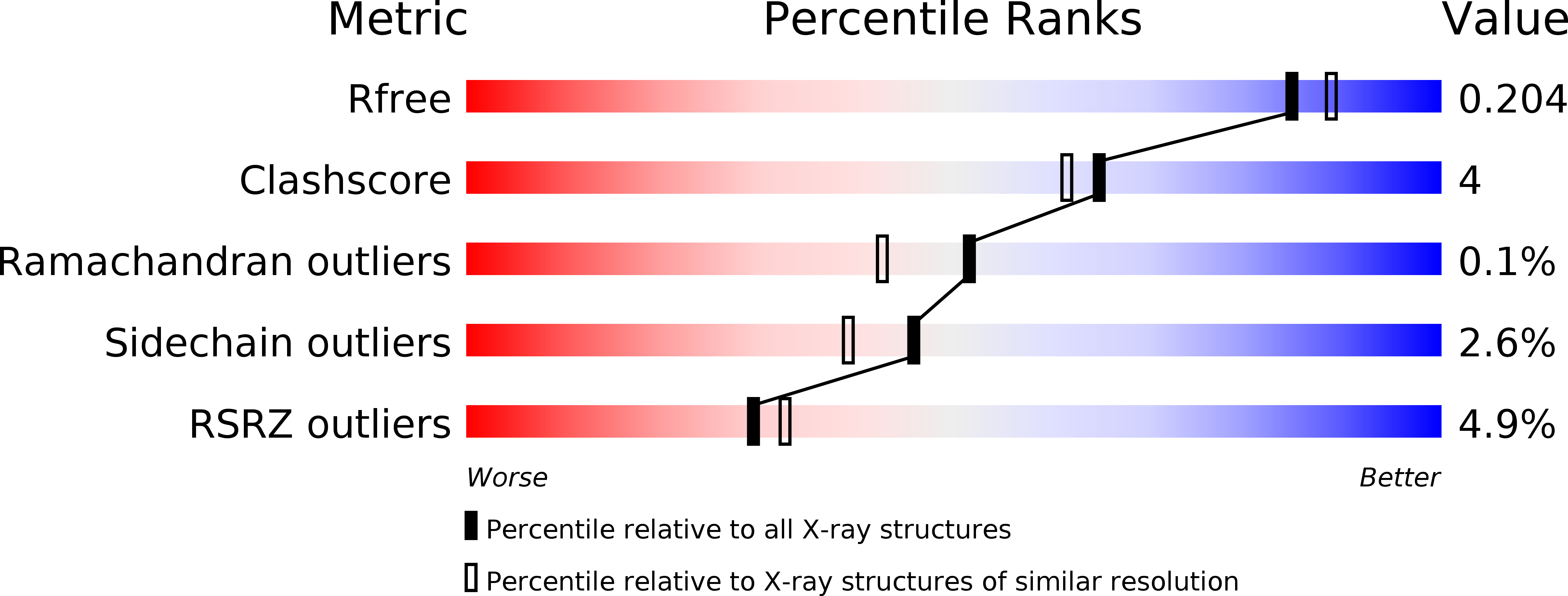
Resolution:
1.90 Å
R-Value Free:
0.20
R-Value Work:
0.17
R-Value Observed:
0.17
Space Group:
P 65 2 2