2IBN image
Deposition Date 2006-09-11
Release Date 2006-10-17
Last Version Date 2024-10-30
Entry Detail
PDB ID:
2IBN
Keywords:
Title:
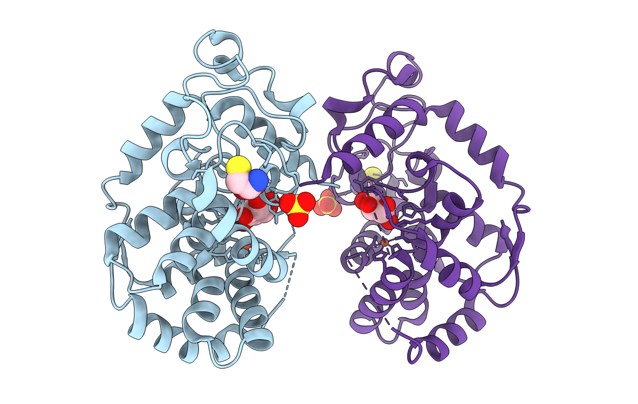
Crystal structure of Human myo-Inositol Oxygenase (MIOX)
Biological Source:
Source Organism:
Homo sapiens (Taxon ID: 9606)
Host Organism:
Method Details:
Experimental Method:
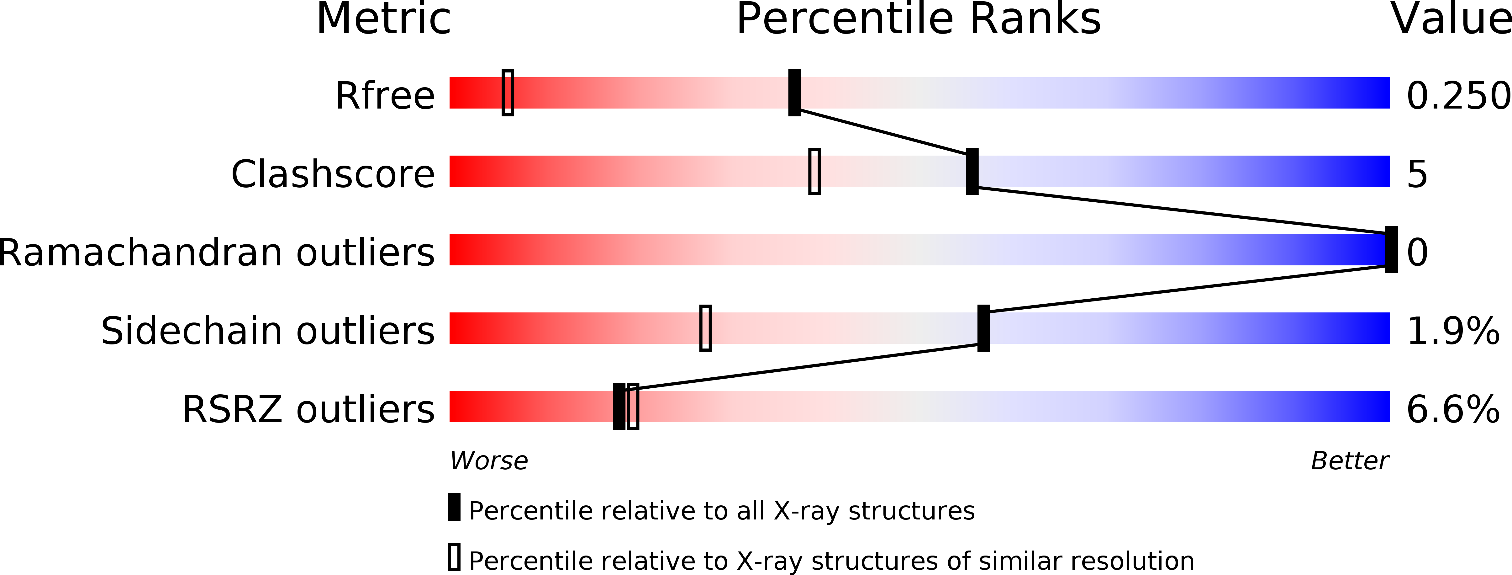
Resolution:
1.50 Å
R-Value Free:
0.24
R-Value Work:
0.20
R-Value Observed:
0.20
Space Group:
C 1 2 1
Macromolecular Entities
Structures with similar UniProt ID
Protein Blast
Polymer Type:polypeptide(L)
Molecule:Inositol oxygenase
Gene (Uniprot):MIOX
Chain IDs:A, B
Chain Length:250
Number of Molecules:2
Biological Source:Homo sapiens
Modified Residue
Compound ID Chain ID Parent Comp ID Details 2D Image
MSE A MET SELENOMETHIONINE
Primary Citation
Structural and Biophysical Characterization of Human myo-Inositol Oxygenase
J.Biol.Chem. 283 15209 15216 (2008)
PMID: 18364358 DOI: 10.1074/jbc.M800348200

Abstact

Altered inositol metabolism is implicated in a number of diabetic complications. The first committed step in mammalian inositol catabolism is performed by myo-inositol oxygenase (MIOX), which catalyzes a unique four-electron dioxygen-dependent ring cleavage of myo-inositol to D-glucuronate. Here, we present the crystal structure of human MIOX in complex with myo-inosose-1 bound in a terminal mode to the MIOX diiron cluster site. Furthermore, from biochemical and biophysical results from N-terminal deletion mutagenesis we show that the N terminus is important, through coordination of a set of loops covering the active site, in shielding the active site during catalysis. EPR spectroscopy of the unliganded enzyme displays a two-component spectrum that we can relate to an open and a closed active site conformation. Furthermore, based on site-directed mutagenesis in combination with biochemical and biophysical data, we propose a novel role for Lys(127) in governing access to the diiron cluster.

Legend

Protein

Chemical

Disease

Primary Citation of related structures