
Deposition Date
2006-09-11
Release Date
2007-04-03
Last Version Date
2023-10-25
Entry Detail
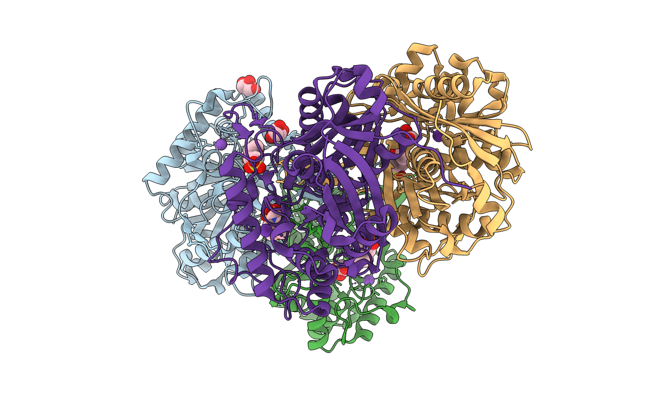
PDB ID:
2IB8
Keywords:
Title:
Crystallographic and kinetic studies of human mitochondrial acetoacetyl-CoA thiolase (T2): the importance of potassium and chloride for its structure and function
Biological Source:
Source Organism(s):
Homo sapiens (Taxon ID: 9606)
Expression System(s):
Method Details:
Experimental Method:
Resolution:
1.85 Å
R-Value Free:
0.20
R-Value Work:
0.16
R-Value Observed:
0.16
Space Group:
P 1 21 1
