
Deposition Date
1998-03-13
Release Date
1998-11-18
Last Version Date
2024-11-20
Entry Detail
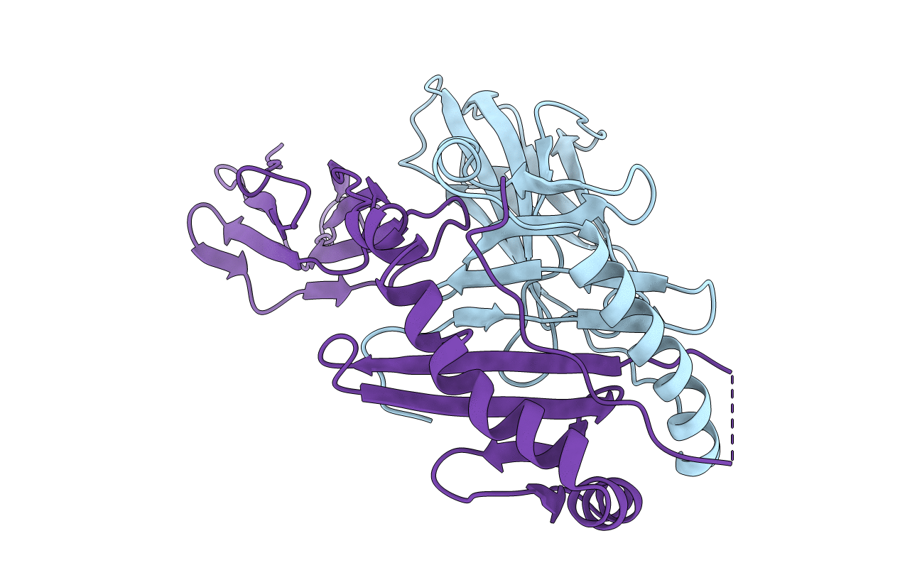
PDB ID:
2IAD
Keywords:
Title:
CLASS II MHC I-AD IN COMPLEX WITH AN INFLUENZA HEMAGGLUTININ PEPTIDE 126-138
Biological Source:
Source Organism(s):
Mus musculus (Taxon ID: 10090)
Expression System(s):
Method Details:
Experimental Method:
Resolution:
2.40 Å
R-Value Free:
0.30
R-Value Work:
0.25
R-Value Observed:
0.25
Space Group:
C 1 2 1
