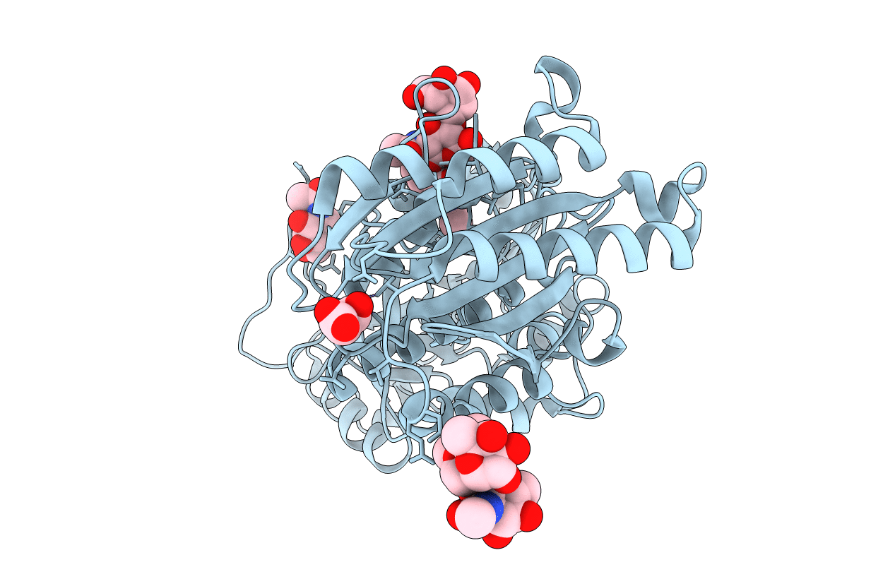
2I6Q image
Entry Detail
PDB ID:
2I6Q
Keywords:
Title:
Complement component C2a
Biological Source:
Source Organism:
Host Organism:
PDB Version:
Deposition Date:
2006-08-29
Release Date:
2006-10-17
Method Details:
Experimental Method:
Resolution:
2.10 Å
R-Value Free:
0.24
R-Value Work:
0.18
Space Group:
P 1 21 1
Macromolecular Entities
Polymer Type:polypeptide(L)
Description:Complement C2a fragment
Chain IDs:A
Chain Length:517
Number of Molecules:1
Biological Source:Homo sapiens
Modified Residue
Compound ID Chain ID Parent Comp ID Details 2D Image
ASN A ASN GLYCOSYLATION SITE
Primary Citation
Structure of complement component c2a: implications for convertase formation and substrate binding.
Structure 14 1587 1597 (2006)
PMID: 17027507 DOI: 10.1016/j.str.2006.08.008

Abstact

C2a provides the catalytic center to the convertase complexes of the classical and lectin-binding pathways of complement activation. We determined two crystal structures of full-length C2a, with and without a pseudo ligand bound. Both structures reveal a near-active conformation of the catalytic center of the serine protease domains, while the von Willebrand factor A-type domains display an intermediate activation state of helix alpha7 with an open, activated metal-ion-dependent adhesion site. The open adhesion site likely serves to enhance the affinity for the ligand C4b, similar to "inside-out" signaling in integrins. Surprisingly, the N-terminal residues of C2a are buried in a crevice near helix alpha7, indicative of a structural switch between C2 and C2a. Extended loops on the protease domain possibly envelop the protruding anaphylatoxin domain of the substrate C3. Together with a putative substrate-induced completion of the oxyanion hole, this may contribute to the high substrate specificity of the convertases.

Legend

Protein

Chemical

Disease

Primary Citation of related structures