2I5W image
Deposition Date 2006-08-26
Release Date 2006-10-17
Last Version Date 2024-02-21
Entry Detail
PDB ID:
2I5W
Title:
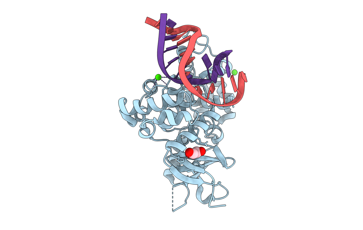
Structure of hOGG1 crosslinked to DNA sampling a normal G adjacent to an oxoG
Biological Source:
Source Organism:
Homo sapiens (Taxon ID: 9606)
Method Details:
Experimental Method:
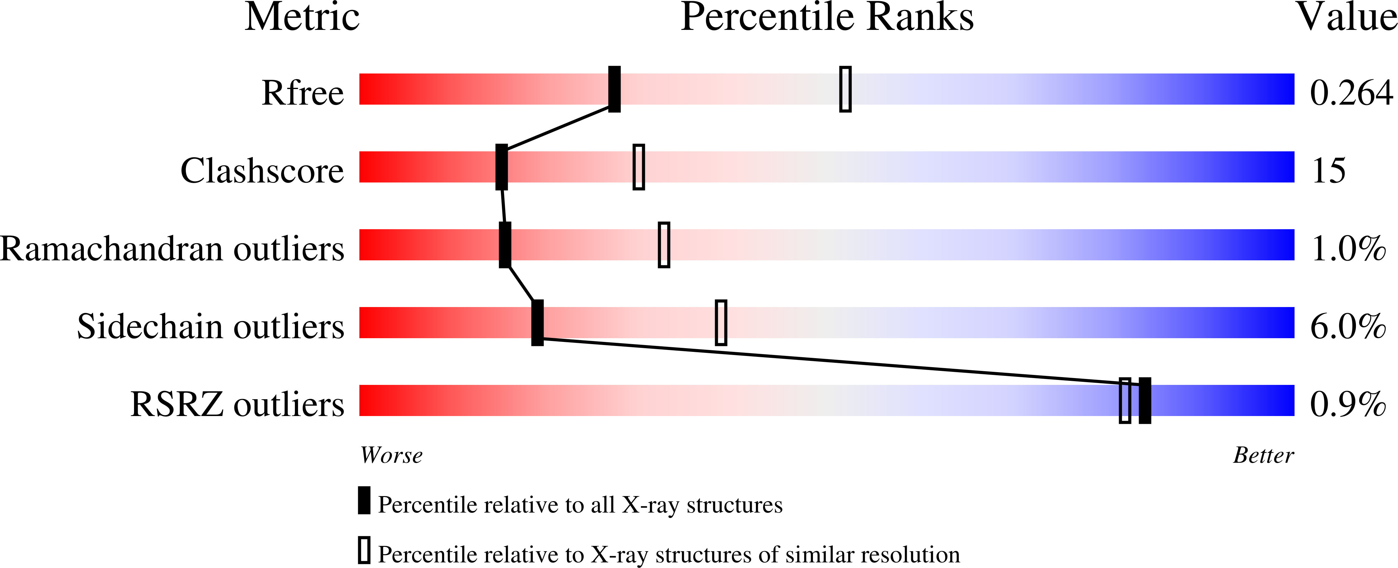
Resolution:
2.60 Å
R-Value Free:
0.26
R-Value Work:
0.22
Space Group:
P 65 2 2
Macromolecular Entities
Structures with similar UniProt ID
Protein Blast
Polymer Type:polypeptide(L)
Molecule:N-glycosylase/DNA lyase
Gene (Uniprot):OGG1
Mutagens:N149C
Chain IDs:C (auth: A)
Chain Length:315
Number of Molecules:1
Biological Source:Homo sapiens
Polymer Type:polydeoxyribonucleotide
Molecule:5'-D(*GP*GP*TP*AP*GP*AP*CP*CP*TP*GP*GP*A)-3'
Chain IDs:A (auth: E)
Chain Length:12
Number of Molecules:1
Biological Source:
Polymer Type:polydeoxyribonucleotide
Molecule:5'-D(P*CP*CP*AP*GP*(G42)P*TP*CP*TP*AP*C)-3'
Chain IDs:B (auth: F)
Chain Length:10
Number of Molecules:1
Biological Source:
Modified Residue
Compound ID Chain ID Parent Comp ID Details 2D Image
8OG B DG ?
Primary Citation
A nucleobase lesion remodels the interaction of its normal neighbor in a DNA glycosylase complex.
Proc.Natl.Acad.Sci.Usa 103 15020 15025 (2006)
PMID: 17015827 DOI: 10.1073/pnas.0603644103

Abstact

How DNA glycosylases search through millions of base pairs and discriminate between rare sites of damage and otherwise undamaged bases is poorly understood. Even less understood are the details of the structural states arising from DNA glycosylases interacting with undamaged DNA. Recognizing the mutagenic lesion 7,8-dihydro-8-oxoguanine (8-oxoguanine, oxoG) represents an especially formidable challenge, because this oxidized nucleobase differs by only two atoms from its normal counterpart, guanine (G), and buried in the structure of naked B-form DNA, oxoG and G are practically indistinguishable from each other. We have used disulfide cross-linking technology to capture a human oxoG repair protein, 8-oxoguanine DNA glycosylase I (hOGG1) sampling an undamaged G:C base pair located adjacent to an oxoG:C base pair in DNA. The x-ray structure of the trapped complex reveals that the presence of the 8-oxoG drastically changes the local conformation of the extruded G. The extruded but intrahelical state of the G in this structure offers a view of an early intermediate in the base-extrusion pathway.

Legend

Protein

Chemical

Disease

Primary Citation of related structures