2I5P image
Deposition Date 2006-08-25
Release Date 2006-09-12
Last Version Date 2023-08-30
Entry Detail
PDB ID:
2I5P
Keywords:
Title:
Crystal structure of glyceraldehyde-3-phosphate dehydrogenase isoform 1 from K. marxianus
Biological Source:
Source Organism:
Host Organism:
Method Details:
Experimental Method:
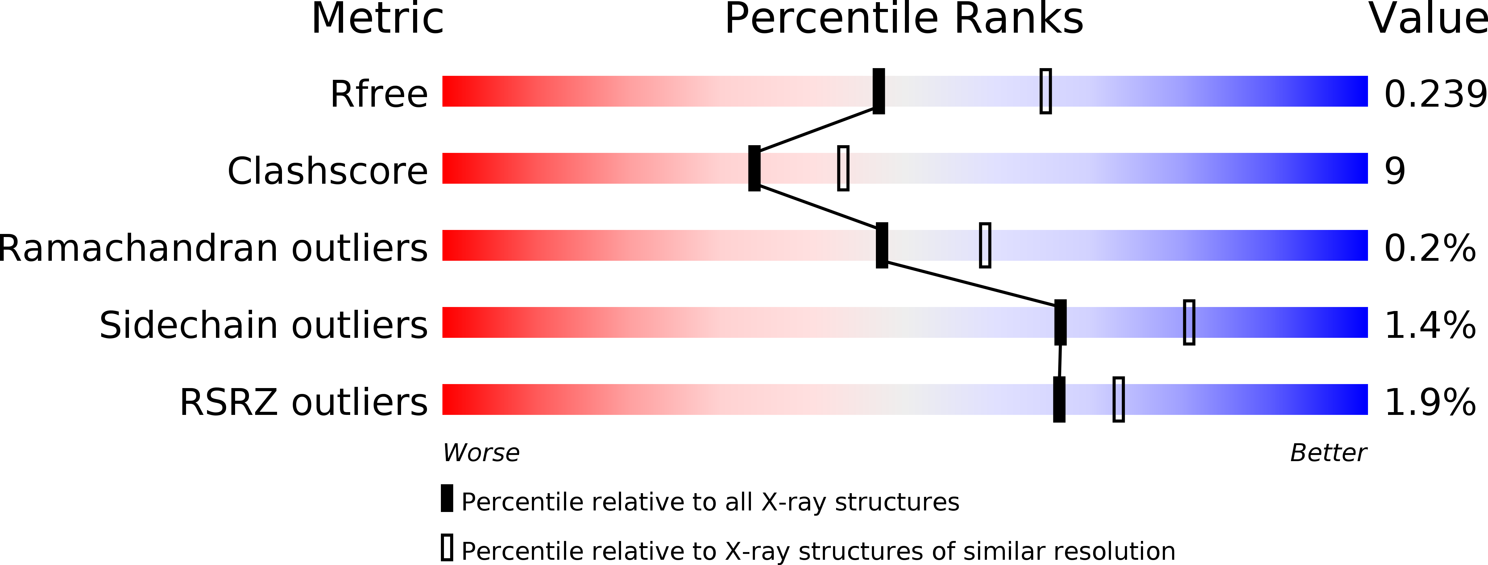
Resolution:
2.30 Å
R-Value Free:
0.24
R-Value Work:
0.21
R-Value Observed:
0.21
Space Group:
P 41 21 2
Macromolecular Entities
Polymer Type:polypeptide(L)
Molecule:Glyceraldehyde-3-phosphate dehydrogenase 1
Gene (Uniprot):GAP1
Chain IDs:A (auth: O), B (auth: P)
Chain Length:342
Number of Molecules:2
Biological Source:Kluyveromyces marxianus
Primary Citation
The Crystal and Solution Structures of Glyceraldehyde-3-phosphate Dehydrogenase Reveal Different Quaternary Structures.
J.Biol.Chem. 281 33433 33440 (2006)
PMID: 16963457 DOI: 10.1074/jbc.M605267200

Abstact

The presence of an isoform of glyceraldehyde-3-phosphate dehydrogenase (kmGAPDH1p) associated with the cell wall of a flocculent strain of Kluyveromyces marxianus was the first report of a non-cytosolic localization of a glycolytic enzyme, but the mechanism by which the protein is transported to the cell surface is not known. To identify structural features that could account for the multiple localizations of the protein, the three-dimensional structure of kmGAPDH1p was determined by x-ray crystallography and small angle x-ray scattering. The x-ray crystallographic structure of kmGAPDH1p revealed a dimer, although all GAPDH homologs studied thus far have a tetrameric structure with 222 symmetry. Interestingly, the structure of kmGAPDH1p in solution revealed a tetramer with a 70 degrees tilt angle between the dimers. Moreover, the separation between the centers of the dimers composing the kmGAPDH1p tetramer diminished from 34 to 30 A upon NAD(+) binding, this latter value being similar to the observed in the crystallographic models of GAPDH homologs. The less compact structure of apo-kmGAPDH1p could already be the first image of the transition intermediate between the tetramer observed in solution and the dimeric form found in the crystal structure, which we postulate to exist in vivo because of the protein's multiple subcellular localizations in this yeast species.

Legend

Protein

Chemical

Disease

Primary Citation of related structures