2I3V image
Deposition Date 2006-08-21
Release Date 2006-10-17
Last Version Date 2024-10-30
Entry Detail
PDB ID:
2I3V
Title:
Measurement of conformational changes accompanying desensitization in an ionotropic glutamate receptor: Structure of G725C mutant
Biological Source:
Source Organism:
Host Organism:
Method Details:
Experimental Method:
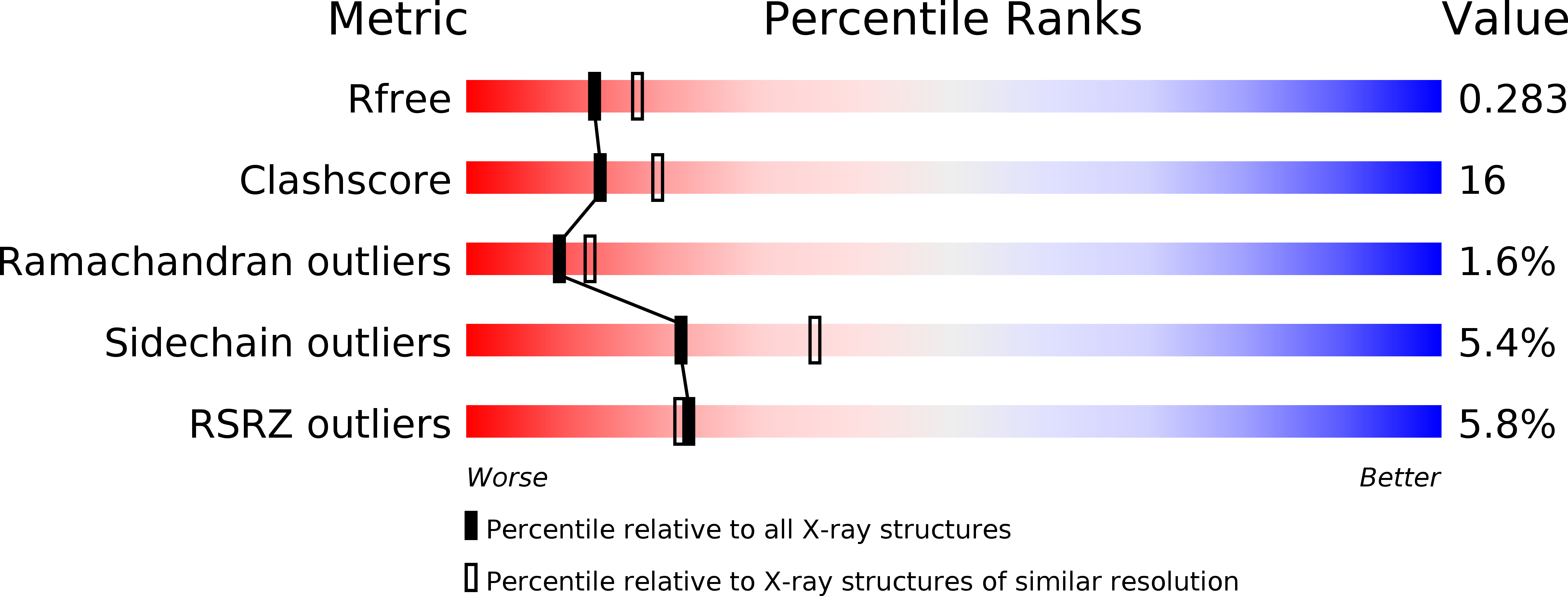
Resolution:
2.40 Å
R-Value Free:
0.28
R-Value Work:
0.23
R-Value Observed:
0.23
Space Group:
P 21 21 21
Macromolecular Entities
Polymer Type:polypeptide(L)
Molecule:Glutamate receptor 2
Gene (Uniprot):Gria2
Chain IDs:A, B, C, D
Chain Length:259
Number of Molecules:4
Biological Source:Rattus norvegicus
Primary Citation
Measurement of Conformational Changes accompanying Desensitization in an Ionotropic Glutamate Receptor.
Cell(Cambridge,Mass.) 127 85 97 (2006)
PMID: 17018279 DOI: 10.1016/j.cell.2006.08.037

Abstact

The canonical conformational states occupied by most ligand-gated ion channels, and many cell-surface receptors, are the resting, activated, and desensitized states. While the resting and activated states of multiple receptors are well characterized, elaboration of the structural properties of the desensitized state, a state that is by definition inactive, has proven difficult. Here we use electrical, chemical, and crystallographic experiments on the AMPA-sensitive GluR2 receptor, defining the conformational rearrangements of the agonist binding cores that occur upon desensitization of this ligand-gated ion channel. These studies demonstrate that desensitization involves the rupture of an extensive interface between domain 1 of 2-fold related glutamate-binding core subunits, compensating for the ca. 21 degrees of domain closure induced by glutamate binding. The rupture of the domain 1 interface allows the ion channel to close and thereby provides a simple explanation to the long-standing question of how agonist binding is decoupled from ion channel gating upon receptor desensitization.

Legend

Protein

Chemical

Disease

Primary Citation of related structures