Deposition Date
2006-08-17
Release Date
2006-10-17
Last Version Date
2024-10-30
Method Details:
Experimental Method:
Resolution:
4.15 Å
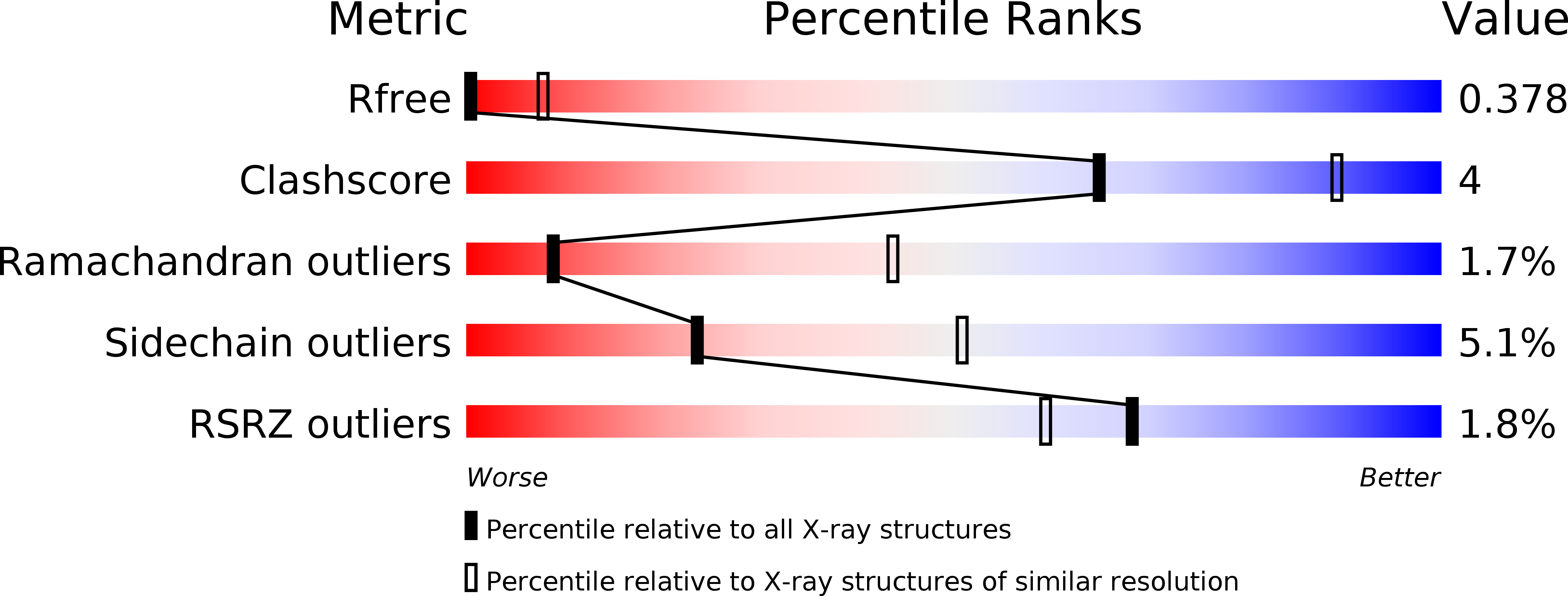
R-Value Free:
0.38
R-Value Work:
0.37
R-Value Observed:
0.37
Space Group:
P 31 1 2