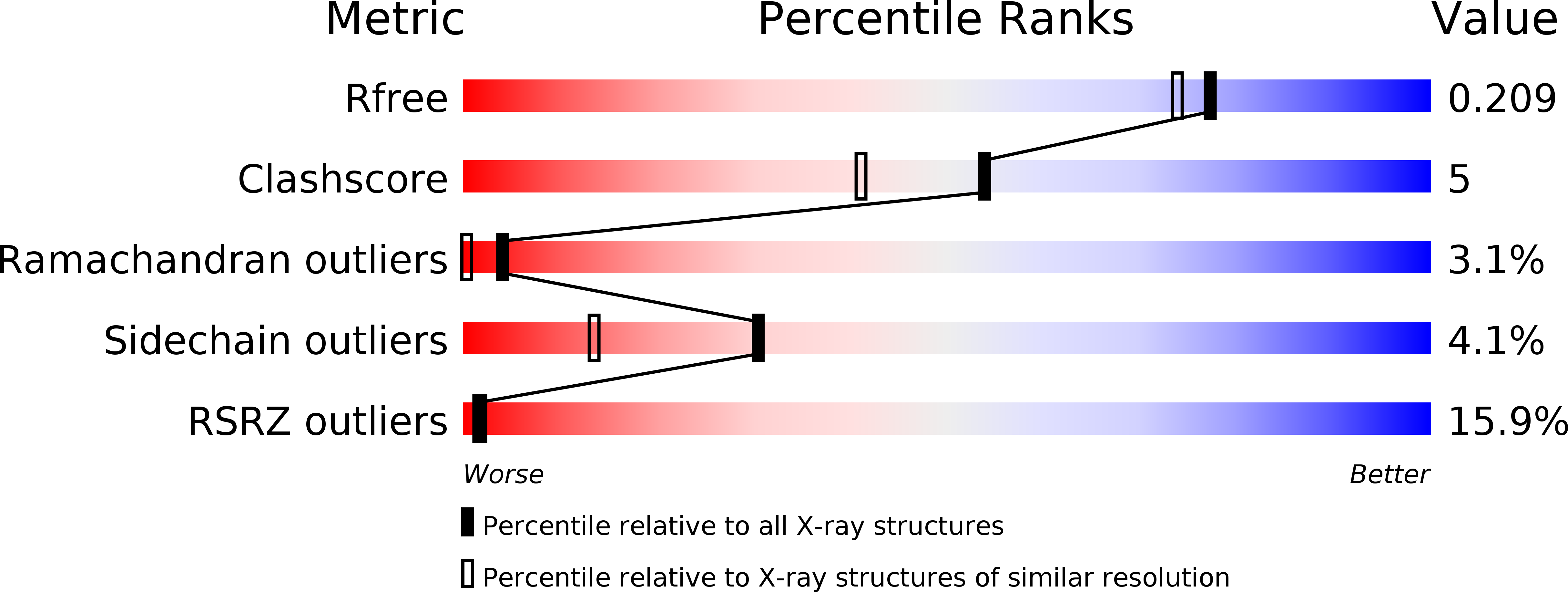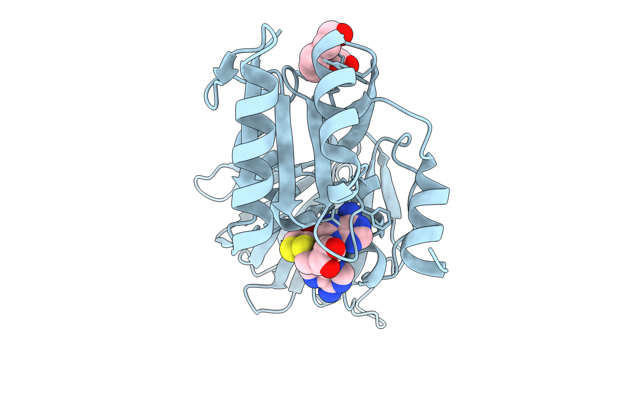
2I2C image
Deposition Date 2006-08-16
Release Date 2007-08-07
Last Version Date 2023-08-30
Entry Detail
PDB ID:
2I2C
Keywords:
Title:
Crystal structure of LmNADK1
Biological Source:
Source Organism:
Host Organism:
Method Details:
Experimental Method:
Resolution:
1.85 Å
R-Value Free:
0.21
R-Value Work:
0.19
R-Value Observed:
0.19
Space Group:
I 2 2 2
Macromolecular Entities
Polymer Type:polypeptide(L)
Molecule:Probable inorganic polyphosphate/ATP-NAD kinase 1
Gene (Uniprot):nadK1
Chain IDs:A
Chain Length:272
Number of Molecules:1
Biological Source:Listeria monocytogenes EGD-e
Primary Citation
NAD kinases use substrate-assisted catalysis for specific recognition of NAD.
J.Biol.Chem. 282 33925 33934 (2007)
PMID: 17686780 DOI: 10.1074/jbc.M701394200

Abstact

Here we describe the crystal structures of the NAD kinase (LmNADK1) from Listeria monocytogenes in complex with its substrate NAD, its product NADP, or two synthesized NAD mimics. We identified one of the NAD mimics, di-adenosine diphosphate, as a new substrate for LmNADK1, whereas we showed that the closely related compound di-5'-thioadenosine is a novel non-natural inhibitor for this enzyme. These structures suggest a mechanism involving substrate-assisted catalysis. Indeed, sequence/structure comparison and directed mutagenesis have previously shown that NAD kinases (NADKs) and the distantly related 6-phosphofructokinases share the same catalytically important GGDGT motif. However, in this study we have shown that these enzymes use the central aspartate of this motif differently. Although this acidic residue chelates the catalytic Mg(2+) ion in 6-phosphofructokinases, it activates the phospho-acceptor (NAD) in NADKs. Sequence/structure comparisons suggest that the role of this aspartate would be conserved in NADKs and the related sphingosine and diacylglycerol kinases.

Legend

Protein

Chemical

Disease

Primary Citation of related structures