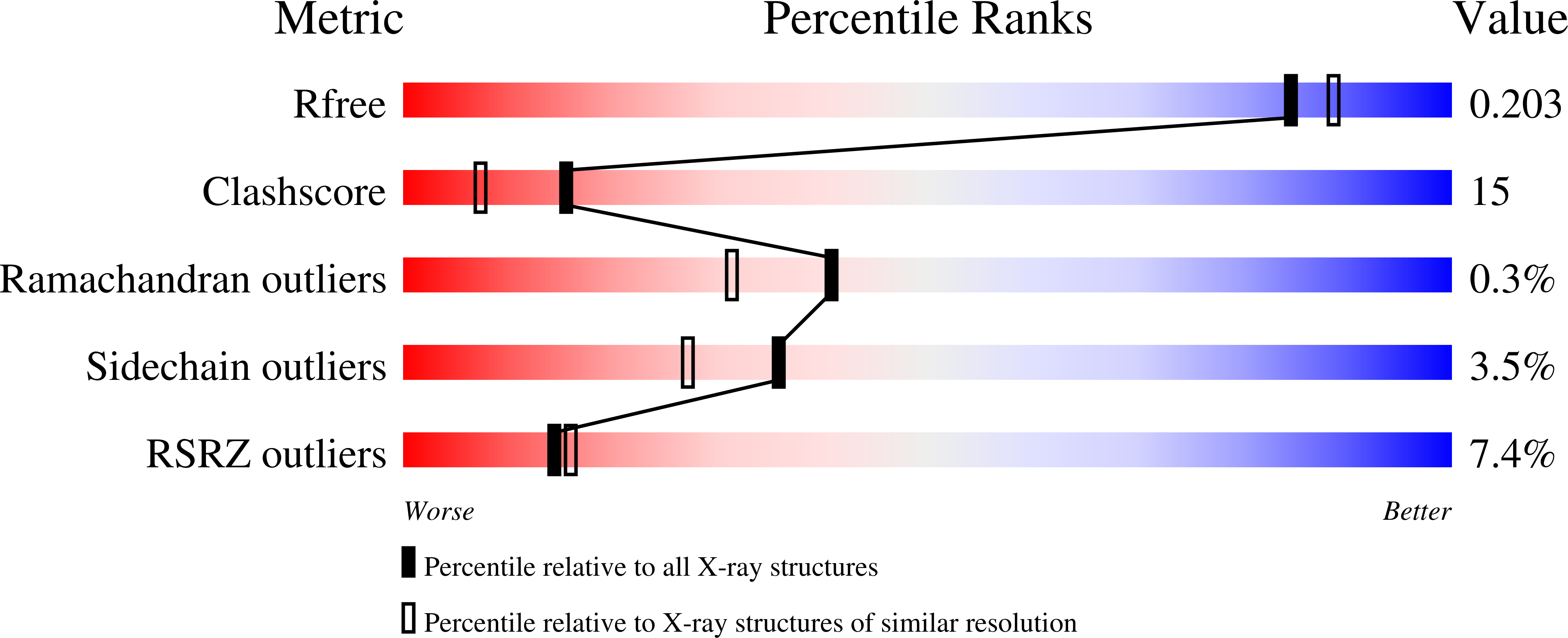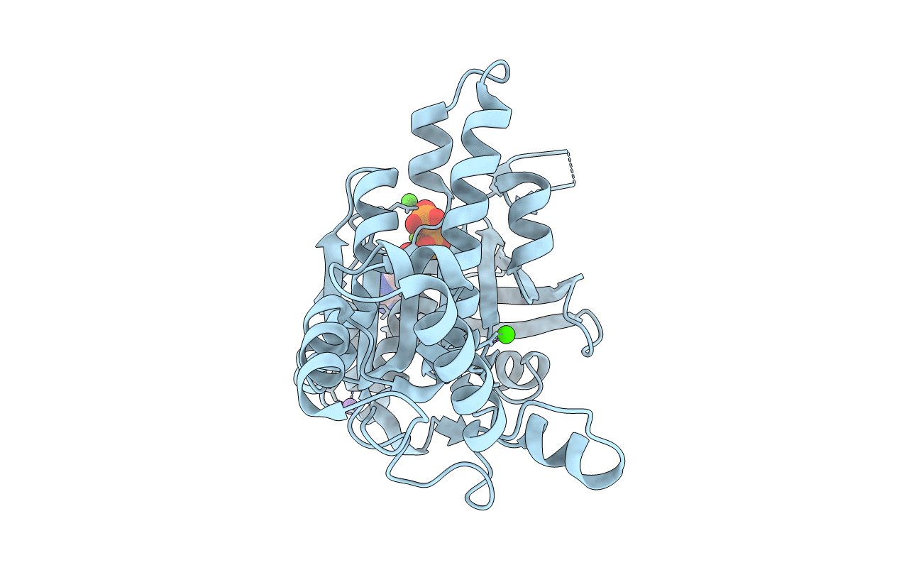
2I1Q image
Deposition Date 2006-08-14
Release Date 2006-10-24
Last Version Date 2023-08-30
Entry Detail
PDB ID:
2I1Q
Keywords:
Title:
RadA Recombinase in complex with Calcium
Biological Source:
Source Organism:
Host Organism:
Method Details:
Experimental Method:
Resolution:
1.90 Å
R-Value Free:
0.21
R-Value Work:
0.19
R-Value Observed:
0.19
Space Group:
P 61
Macromolecular Entities
Polymer Type:polypeptide(L)
Molecule:DNA repair and recombination protein radA
Gene (Uniprot):radA
Mutations:S2G
Chain IDs:A
Chain Length:322
Number of Molecules:1
Biological Source:Methanococcus voltae
Primary Citation
Calcium Stiffens Archaeal Rad51 Recombinase from Methanococcus voltae for Homologous Recombination.
J.Biol.Chem. 281 39380 39387 (2006)
PMID: 17050545 DOI: 10.1074/jbc.M607785200

Abstact

Archaeal RadA or Rad51 recombinases are close homologues of eukaryal Rad51 and DMC1. These and bacterial RecA orthologues play a key role in DNA repair by forming helical nucleoprotein filaments in which a hallmark strand exchange reaction between homologous DNA substrates occurs. Recent studies have discovered the stimulatory role by calcium on human and yeast recombinases. Here we report that the strand exchange activity but not the ATPase activity of an archaeal RadA/Rad51 recombinase from Methanococcus voltae (MvRadA) is also subject to calcium stimulation. Crystallized MvRadA filaments in the presence of CaCl(2) resemble that of the recently reported ATPase active form in the presence of an activating dose of KCl. At the ATPase center, one Ca(2+) ion takes the place of two K(+) ions in the K(+)-bound form. The terminal phosphate of the nonhydrolyzable ATP analogue is in a staggered conformation in the Ca(2+)-bound form. In comparison, an eclipsed conformation was seen in the K(+)-bound form. Despite the changes in the ATPase center, both forms harbor largely ordered L2 regions in essentially identical conformations. These data suggest a unified stimulation mechanism by potassium and calcium because of the existence of a conserved ATPase center promiscuous in binding cations.

Legend

Protein

Chemical

Disease

Primary Citation of related structures