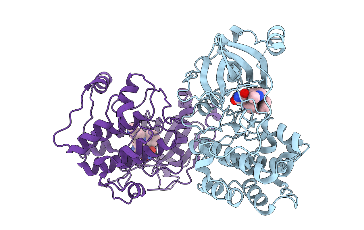
2I0E image
Entry Detail
PDB ID:
2I0E
Keywords:
Title:
Structure of catalytic domain of human protein kinase C beta II complexed with a bisindolylmaleimide inhibitor
Biological Source:
Source Organism:
Host Organism:
PDB Version:
Deposition Date:
2006-08-10
Release Date:
2006-11-14
Method Details:
Experimental Method:
Resolution:
2.60 Å
R-Value Free:
0.28
R-Value Work:
0.23
R-Value Observed:
0.23
Space Group:
P 21 21 2
Macromolecular Entities
Polymer Type:polypeptide(L)
Description:Protein Kinase C-beta II
Chain IDs:A, B
Chain Length:353
Number of Molecules:2
Biological Source:Homo sapiens
Modified Residue
Compound ID Chain ID Parent Comp ID Details 2D Image
SEP A SER PHOSPHOSERINE
TPO A THR PHOSPHOTHREONINE
Ligand Molecules
Primary Citation
Structure of the Catalytic Domain of Human Protein Kinase C beta II Complexed with a Bisindolylmaleimide Inhibitor
Biochemistry 45 13970 13981 (2006)
PMID: 17115692 DOI: 10.1021/bi061128h

Abstact

The conventional protein kinase C isoform, PKCII, is a signaling kinase activated during the hyperglycemic state and has been associated with the development of microvascular abnormalities associated with diabetes. PKCII, therefore, has been identified as a therapeutic target where inhibitors of its kinase activity are being pursued for treatment of microvascular-related diabetic complications. In this report, we describe the crystal structure of the catalytic domain of PKCbetaII complexed with an inhibitor at 2.6 A resolution. The kinase domain of PKCbetaII was cleaved and purified from full-length PKCbetaII expressed in baculovirus-infected insect cells. The overall kinase domain structure follows the classical bilobal fold and is in its fully activated conformation with three well-defined phosphorylated residues: Thr-500, Thr-641, and Ser-660. Different from the crystal structures of nonconventional PKC isoforms, the C-terminus of the PKCbetaII catalytic domain is almost fully ordered and features a novel alpha helix in the turn motif. An ATP-competitive inhibitor, 2-methyl-1H-indol-3-yl-BIM-1, was crystallized with the PKCbetaII catalytic domain as a dimer of two enzyme-inhibitor complexes. The bound inhibitor adopts a nonplanar conformation in the ATP-binding site, with the kinase domain taking on an intermediate, open conformation. This PKCbetaII-inhibitor complex represents the first structural description of any conventional PKC kinase domain. Given the pathogenic role of PKCbetaII in the development of diabetic complications, this structure can serve as a template for the rational design of inhibitors as potential therapeutic agents.

Legend

Protein

Chemical

Disease

Primary Citation of related structures