Deposition Date
2006-08-08
Release Date
2006-08-29
Last Version Date
2024-02-21
Entry Detail
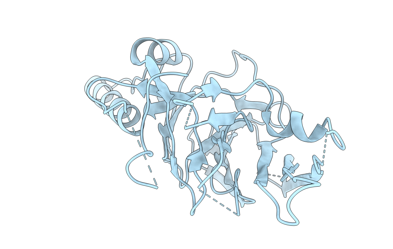
PDB ID:
2HZ6
Keywords:
Title:
The crystal structure of human IRE1-alpha luminal domain
Biological Source:
Source Organism(s):
Homo sapiens (Taxon ID: 9606)
Expression System(s):
Method Details:
Experimental Method:
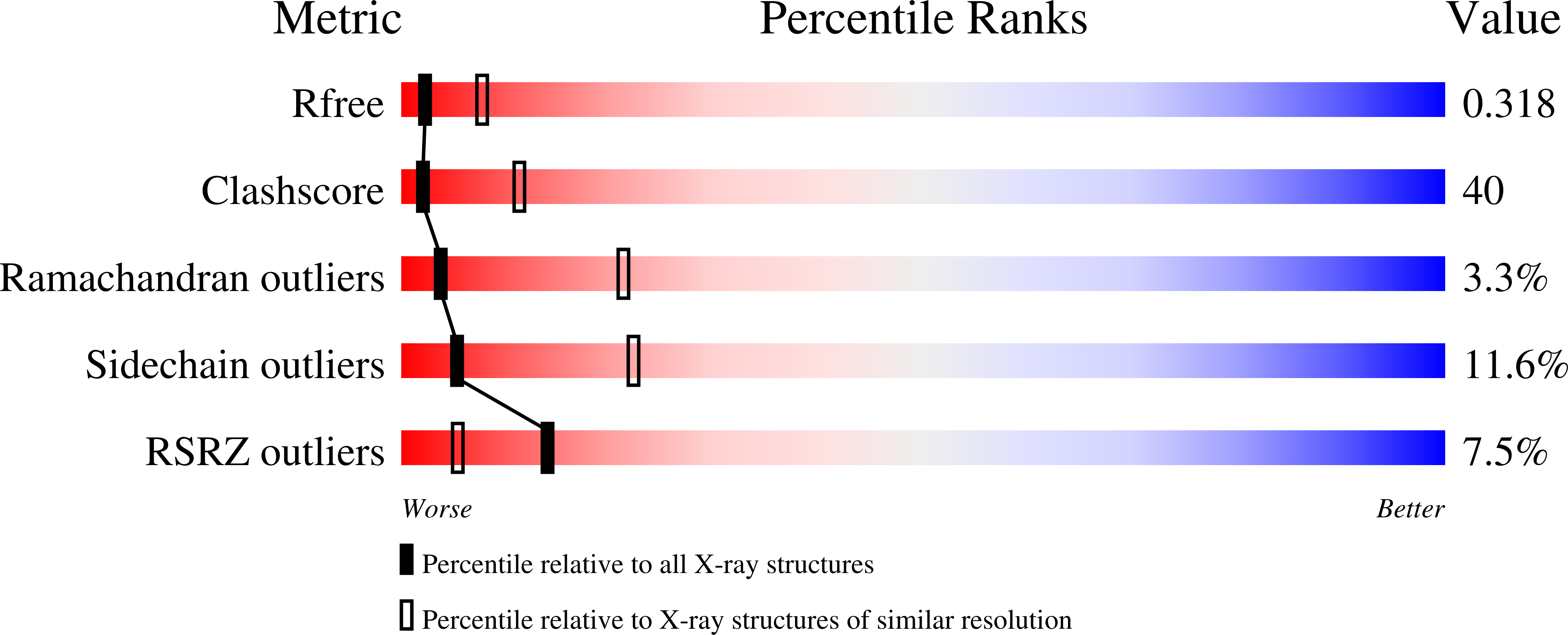
Resolution:
3.10 Å
R-Value Free:
0.31
R-Value Work:
0.26
R-Value Observed:
0.26
Space Group:
P 65 2 2