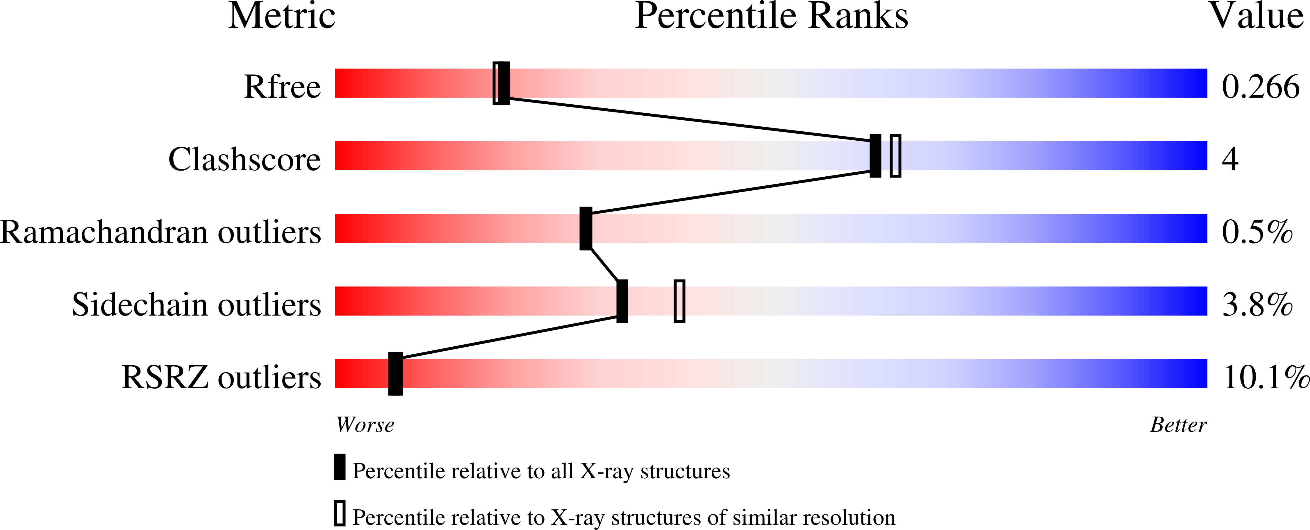
2HYO image
Deposition Date 2006-08-07
Release Date 2006-12-26
Last Version Date 2024-02-21
Entry Detail
PDB ID:
2HYO
Keywords:
Title:
Crystal structure of Rv0805 N97A mutant
Biological Source:
Source Organism:
Method Details:
Experimental Method:
Resolution:
2.25 Å
R-Value Free:
0.26
R-Value Work:
0.21
R-Value Observed:
0.31
Space Group:
P 21 21 2
Macromolecular Entities
Structures with similar UniProt ID
Protein Blast
Polymer Type:polypeptide(L)
Molecule:Rv0805N97A
Mutagens:N97A
Chain IDs:A
Chain Length:280
Number of Molecules:1
Biological Source:Mycobacterium tuberculosis
Primary Citation
Structural and Biochemical Analysis of the Rv0805 Cyclic Nucleotide Phosphodiesterase from Mycobacterium tuberculosis.
J.Mol.Biol. 365 211 225 (2007)
PMID: 17059828 DOI: 10.1016/j.jmb.2006.10.005

Abstact

Cyclic nucleotide monophosphate (cNMP) hydrolysis in bacteria and eukaryotes is brought about by distinct cNMP phosphodiesterases (PDEs). Since these enzymes differ in amino acid sequence and properties, they have evolved by convergent evolution. Cyclic NMP PDEs cleave cNMPs to NMPs, and the Rv0805 gene product is, to date, the only identifiable cNMP PDE in the genome of Mycobacterium tuberculosis. We have shown that Rv0805 is a cAMP/cGMP dual specificity PDE, and is unrelated in amino acid sequence to the mammalian cNMP PDEs. Rv0805 is a dimeric, Fe(3+)-Mn(2+) binuclear PDE, and mutational analysis demonstrated that the active site metals are co-ordinated by conserved aspartate, histidine and asparagine residues. We report here the structure of the catalytic core of Rv0805, which is distantly related to the calcineurin-like phosphatases. The crystal structure of the Rv0805 dimer shows that the active site metals contribute to dimerization and thus play an additional structural role apart from their involvement in catalysis. We also present the crystal structures of the Asn97Ala mutant protein that lacks one of the Mn(2+) co-ordinating residues as well as the Asp66Ala mutant that has a compromised cAMP hydrolytic activity, providing a structural basis for the catalytic properties of these mutant proteins. A molecule of phosphate is bound in a bidentate manner at the active site of the Rv0805 wild-type protein, and cacodylate occupies a similar position in the crystal structure of the Asp66Ala mutant protein. A unique substrate binding pocket in Rv0805 was identified by computational docking studies, and the role of the His140 residue in interacting with cAMP was validated through mutational analysis. This report on the first structure of a bacterial cNMP PDE thus significantly extends our molecular understanding of cAMP hydrolysis in class III PDEs.

Legend

Protein

Chemical

Disease

Primary Citation of related structures