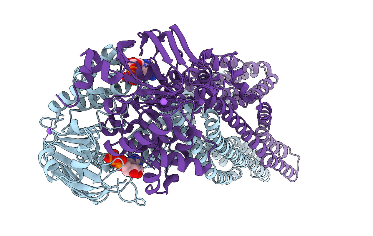
2HYD image
Deposition Date 2006-08-05
Release Date 2006-09-05
Last Version Date 2024-02-21
Entry Detail
PDB ID:
2HYD
Title:
Multidrug ABC transporter SAV1866
Biological Source:
Source Organism:
Host Organism:
Method Details:
Experimental Method:
Resolution:
3.00 Å
R-Value Free:
0.27
R-Value Work:
0.25
R-Value Observed:
0.25
Space Group:
C 1 2 1
Macromolecular Entities
Structures with similar UniProt ID
Protein Blast
Polymer Type:polypeptide(L)
Molecule:ABC transporter homolog
Gene (Uniprot):SAV1866
Chain IDs:A, B
Chain Length:578
Number of Molecules:2
Biological Source:Staphylococcus aureus
Primary Citation
Structure of a bacterial multidrug ABC transporter.
Nature 443 180 185 (2006)
PMID: 16943773 DOI: 10.1038/nature05155

Abstact

Multidrug transporters of the ABC family facilitate the export of diverse cytotoxic drugs across cell membranes. This is clinically relevant, as tumour cells may become resistant to agents used in chemotherapy. To understand the molecular basis of this process, we have determined the 3.0 A crystal structure of a bacterial ABC transporter (Sav1866) from Staphylococcus aureus. The homodimeric protein consists of 12 transmembrane helices in an arrangement that is consistent with cross-linking studies and electron microscopic imaging of the human multidrug resistance protein MDR1, but critically different from that reported for the bacterial lipid flippase MsbA. The observed, outward-facing conformation reflects the ATP-bound state, with the two nucleotide-binding domains in close contact and the two transmembrane domains forming a central cavity--presumably the drug translocation pathway--that is shielded from the inner leaflet of the lipid bilayer and from the cytoplasm, but exposed to the outer leaflet and the extracellular space.

Legend

Protein

Chemical

Disease

Primary Citation of related structures