Deposition Date
2006-08-04
Release Date
2006-10-24
Last Version Date
2024-11-06
Entry Detail
PDB ID:
2HXZ
Keywords:
Title:
Crystal Structure of Cathepsin S in complex with a Nonpeptidic Inhibitor (Hexagonal spacegroup)
Biological Source:
Source Organism(s):
Homo sapiens (Taxon ID: 9606)
Expression System(s):
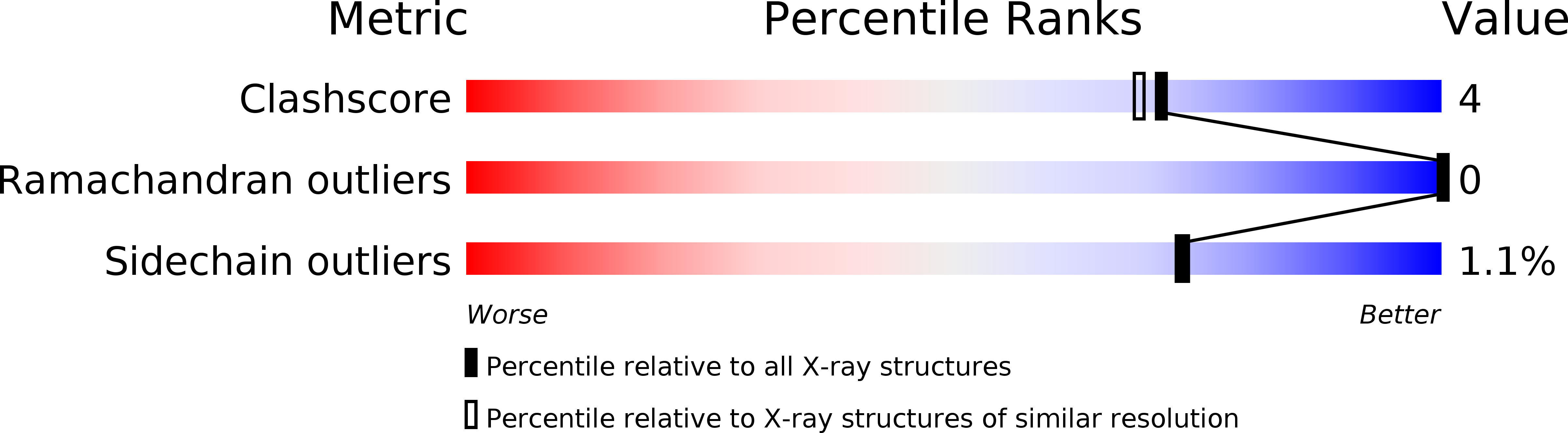
Method Details:
Experimental Method:
Resolution:
1.90 Å
R-Value Free:
0.23
R-Value Work:
0.21
R-Value Observed:
0.22
Space Group:
P 65 2 2