2HXW image
Deposition Date 2006-08-04
Release Date 2007-05-01
Last Version Date 2024-10-16
Entry Detail
PDB ID:
2HXW
Title:
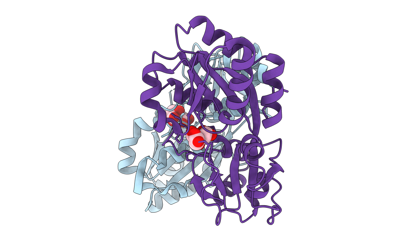
Crystal Structure of Peb3 from Campylobacter jejuni
Biological Source:
Source Organism:
Host Organism:
Method Details:
Experimental Method:
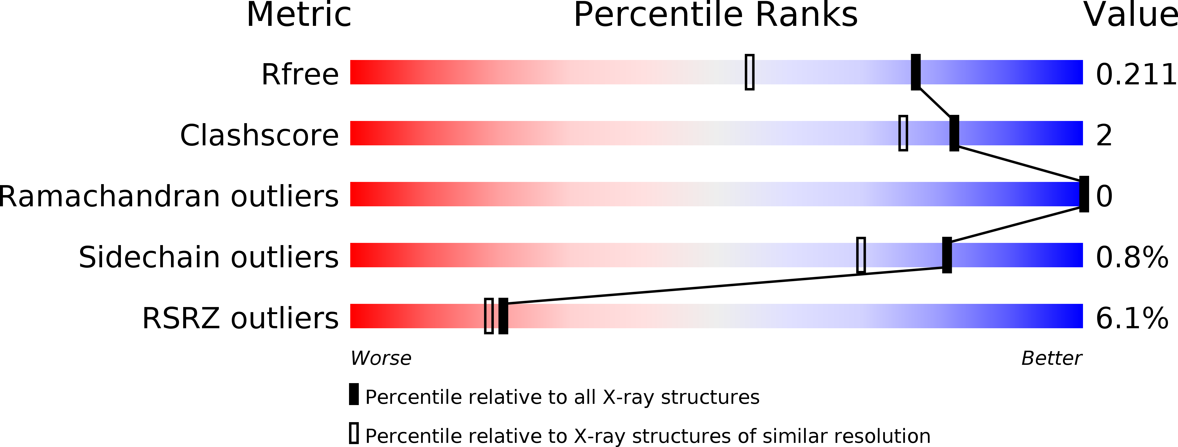
Resolution:
1.60 Å
R-Value Free:
0.21
R-Value Work:
0.18
R-Value Observed:
0.18
Space Group:
P 1 21 1
Macromolecular Entities
Polymer Type:polypeptide(L)
Molecule:Major antigenic peptide PEB3
Chain IDs:A, B
Chain Length:237
Number of Molecules:2
Biological Source:Campylobacter jejuni
Modified Residue
Compound ID Chain ID Parent Comp ID Details 2D Image
MSE A MET SELENOMETHIONINE
Ligand Molecules
Primary Citation
Structural context for protein N-glycosylation in bacteria: The structure of PEB3, an adhesin from Campylobacter jejuni.
Protein Sci. 16 990 995 (2007)
PMID: 17456748 DOI: 10.1110/ps.062737507

Abstact

Campylobacter jejuni is unusual among bacteria in possessing a eukaryotic-like system for N-linked protein glycosylation at Asn residues in sequons of the type Asp/Glu-Xaa-Asn-Xaa-Ser/Thr. However, little is known about the structural context of the glycosylated sequons, limiting the design of novel recombinant glycoproteins. To obtain more information on sequon structure, we have determined the crystal structure of the PEB3 (Cj0289c) dimer. PEB3 has the class II periplasmic-binding protein fold, with each monomer having two domains with a ligand-binding site containing citrate located between them, and overall resembles molybdate- and sulfate-binding proteins. The sequon around Asn90 is located within a surface-exposed loop joining two structural elements. The three key residues are well exposed on the surface; hence, they may be accessible to the PglB oligosaccharyltransferase in the folded state.

Legend

Protein

Chemical

Disease

Primary Citation of related structures