
Deposition Date
2006-08-02
Release Date
2007-01-23
Last Version Date
2023-09-20
Entry Detail
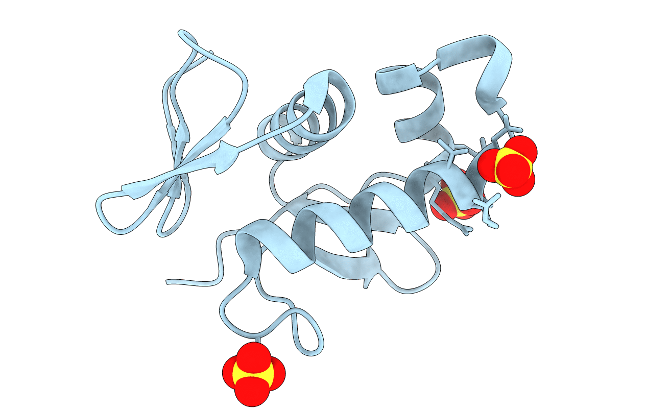
PDB ID:
2HWV
Keywords:
Title:
Crystal structure of an essential response regulator DNA binding domain, VicRc in Enterococcus faecalis, a member of the YycF subfamily.
Biological Source:
Source Organism(s):
Enterococcus faecalis (Taxon ID: 226185)
Expression System(s):
Method Details:
Experimental Method:
Resolution:
1.90 Å
R-Value Free:
0.23
R-Value Work:
0.17
R-Value Observed:
0.18
Space Group:
P 43
