
Deposition Date
2006-08-01
Release Date
2006-11-14
Last Version Date
2023-11-15
Entry Detail
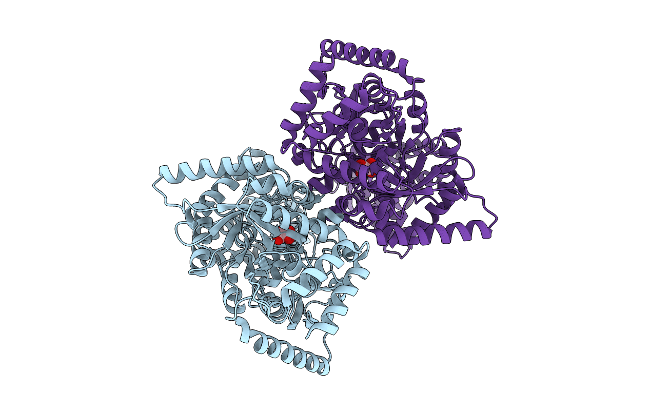
PDB ID:
2HWG
Keywords:
Title:
Structure of phosphorylated Enzyme I of the phosphoenolpyruvate:sugar phosphotransferase system
Biological Source:
Source Organism(s):
Escherichia coli (Taxon ID: 562)
Expression System(s):
Method Details:
Experimental Method:
Resolution:
2.70 Å
R-Value Free:
0.28
R-Value Work:
0.20
R-Value Observed:
0.20
Space Group:
P 21 21 21
