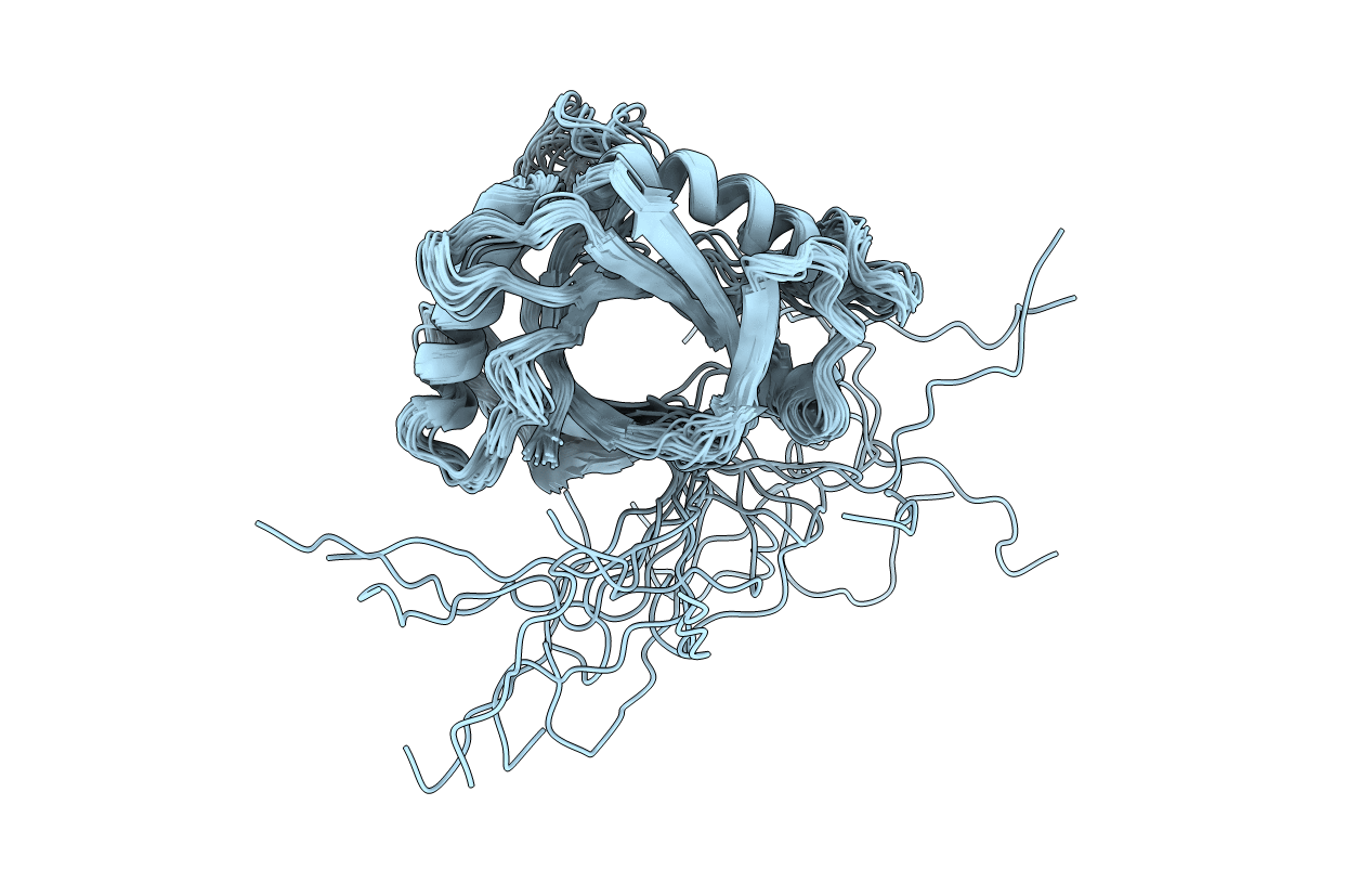
2HVA image
Entry Detail
PDB ID:
2HVA
Title:
Solution Structure of the haem-binding protein p22HBP
Biological Source:
Source Organism:
Host Organism:
PDB Version:
Deposition Date:
2006-07-28
Release Date:
2006-08-08
Method Details:
Experimental Method:
Conformers Calculated:
200
Conformers Submitted:
21
Selection Criteria:
structures with the least restraint violations
Macromolecular Entities
Polymer Type:polypeptide(L)
Description:Heme-binding protein 1
Chain IDs:A
Chain Length:192
Number of Molecules:1
Biological Source:Mus musculus
Ligand Molecules
Primary Citation
A Novel Haem-binding Interface in the 22 kDa Haem-binding Protein p22HBP.
J.Mol.Biol. 362 287 297 (2006)
PMID: 16905148 DOI: 10.1016/j.jmb.2006.07.010

Abstact

The 22 kDa haem-binding protein, p22HBP, is highly expressed in erythropoietic tissues and binds to a range of metallo- and non-metalloporphyrin molecules with similar affinities, suggesting a role in haem regulation or synthesis. We have determined the three-dimensional solution structure of p22HBP and mapped the porphyrin-binding site, which comprises a number of loops and a alpha-helix all located on a single face of the molecule. The structure of p22HBP is related to the bacterial multi-drug resistance protein BmrR, and is the first protein with this fold to be identified in eukaryotes. Strikingly, the porphyrin-binding site in p22HBP is located in a similar position to the drug-binding site of BmrR. These similarities suggest that the broad ligand specificity observed for both BmrR and p22HBP may result from a conserved ligand interaction mechanism. Taken together, these data suggest that the both the fold and its associated function, that of binding to a broad range of small hydrophobic molecules, are ancient, and have been adapted throughout evolution for a variety of purposes.

Legend

Protein

Chemical

Disease

Primary Citation of related structures