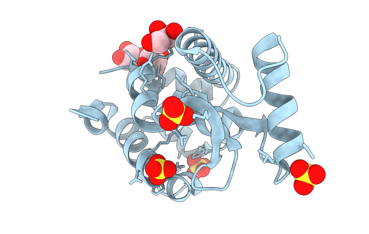
2HUL image
Entry Detail
PDB ID:
2HUL
Keywords:
Title:
Crystal structure of T4 Lysozyme S44C synthetic dimer
Biological Source:
Source Organism:
PDB Version:
Deposition Date:
2006-07-26
Release Date:
2006-10-17
Method Details:
Experimental Method:
Resolution:
1.80 Å
R-Value Free:
0.19
R-Value Work:
0.17
R-Value Observed:
0.17
Space Group:
C 1 2 1
Macromolecular Entities
Polymer Type:polypeptide(L)
Description:Lysozyme
Mutations:S44C
Chain IDs:A
Chain Length:164
Number of Molecules:1
Biological Source:Enterobacteria phage T4
Primary Citation
An approach to crystallizing proteins by synthetic symmetrization.
Proc.Natl.Acad.Sci.Usa 103 16230 16235 (2006)
PMID: 17050682 DOI: 10.1073/pnas.0607674103

Abstact

Previous studies of symmetry preferences in protein crystals suggest that symmetric proteins, such as homodimers, might crystallize more readily on average than asymmetric, monomeric proteins. Proteins that are naturally monomeric can be made homodimeric artificially by forming disulfide bonds between individual cysteine residues introduced by mutagenesis. Furthermore, by creating a variety of single-cysteine mutants, a series of distinct synthetic dimers can be generated for a given protein of interest, with each expected to gain advantage from its added symmetry and to exhibit a crystallization behavior distinct from the other constructs. This strategy was tested on phage T4 lysozyme, a protein whose crystallization as a monomer has been studied exhaustively. Experiments on three single-cysteine mutants, each prepared in dimeric form, yielded numerous novel crystal forms that cannot be realized by monomeric lysozyme. Six new crystal forms have been characterized. The results suggest that synthetic symmetrization may be a useful approach for enlarging the search space for crystallizing proteins.

Legend

Protein

Chemical

Disease

Primary Citation of related structures