2HUG image
Deposition Date 2006-07-26
Release Date 2007-09-18
Last Version Date 2024-05-08
Entry Detail
PDB ID:
2HUG
Keywords:
Title:
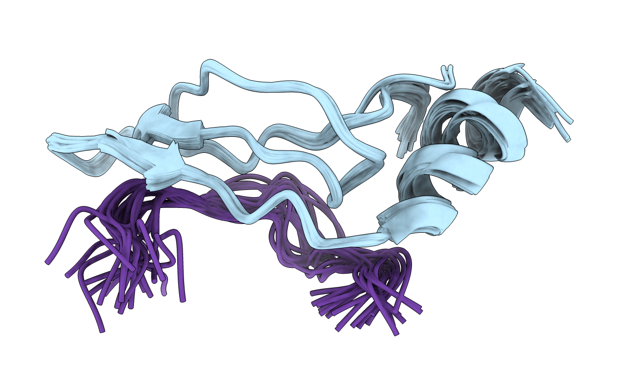
3D Solution Structure of the Chromo-2 Domain of cpSRP43 complexed with cpSRP54 peptide
Biological Source:
Source Organism:
Host Organism:
Method Details:
Experimental Method:
Conformers Calculated:
200
Conformers Submitted:
20
Macromolecular Entities
Polymer Type:polypeptide(L)
Molecule:Signal recognition particle 43 kDa protein, chloroplast
Gene (Uniprot):CAO
Chain IDs:A
Chain Length:57
Number of Molecules:1
Biological Source:Arabidopsis thaliana
Polymer Type:polypeptide(L)
Molecule:Signal recognition particle 54 kDa protein, chloroplast
Gene (Uniprot):FFC
Chain IDs:B
Chain Length:14
Number of Molecules:1
Biological Source:Arabidopsis thaliana
Ligand Molecules
Primary Citation
Assembly of chloroplast signal recognition particle involves structural rearrangement in cpSRP43.
J.Mol.Biol. 381 49 60 (2008)
PMID: 18586266 DOI: 10.1016/j.jmb.2008.05.065

Abstact

Signal recognition particle in chloroplasts (cpSRP) exhibits the unusual ability to bind and target full-length proteins to the thylakoid membrane. Unlike cytosolic SRPs in prokaryotes and eukaryotes, cpSRP lacks an RNA moiety and functions as a heterodimer composed of a conserved 54-kDa guanosine triphosphatase (cpSRP54) and a unique 43-kDa subunit (cpSRP43). Assembly of the cpSRP heterodimer is a prerequisite for post-translational targeting activities and takes place through interactions between chromatin modifier domain 2 (CD2) of cpSRP43 and a unique 10-amino-acid region in cpSRP54 (cpSRP54(pep)). We have used multidimensional NMR spectroscopy and other biophysical methods to examine the assembly and structure of the cpSRP43-cpSRP54 interface. Our data show that CD2 of cpSRP43 binds to cpSRP54(pep) in a 1:1 stoichiometry with an apparent K(d) of approximately 1.06 muM. Steady-state fluorescence and far-UV circular dichroism data suggest that the CD2-cpSRP54(pep) interaction causes significant conformational changes in both CD2 and the peptide. Comparison of the three-dimensional solution structures of CD2 alone and in complex with cpSRP54(pep) shows that significant structural changes are induced in CD2 in order to establish a binding interface contributed mostly by residues in the N-terminal segment of CD2 (Phe5-Val10) and an arginine doublet (Arg536 and Arg537) in the cpSRP54 peptide. Taken together, our results provide new insights into the mechanism of cpSRP assembly and the structural forces that stabilize the functionally critical cpSRP43-cpSRP54 interaction.

Legend

Protein

Chemical

Disease

Primary Citation of related structures