Deposition Date
2006-07-26
Release Date
2006-09-26
Last Version Date
2023-11-15
Entry Detail
PDB ID:
2HUF
Keywords:
Title:
Crystal structure of Aedes aegypti alanine glyoxylate aminotransferase
Biological Source:
Source Organism:
Aedes aegypti (Taxon ID: 7159)
Host Organism:
Method Details:
Experimental Method:
Resolution:
1.75 Å
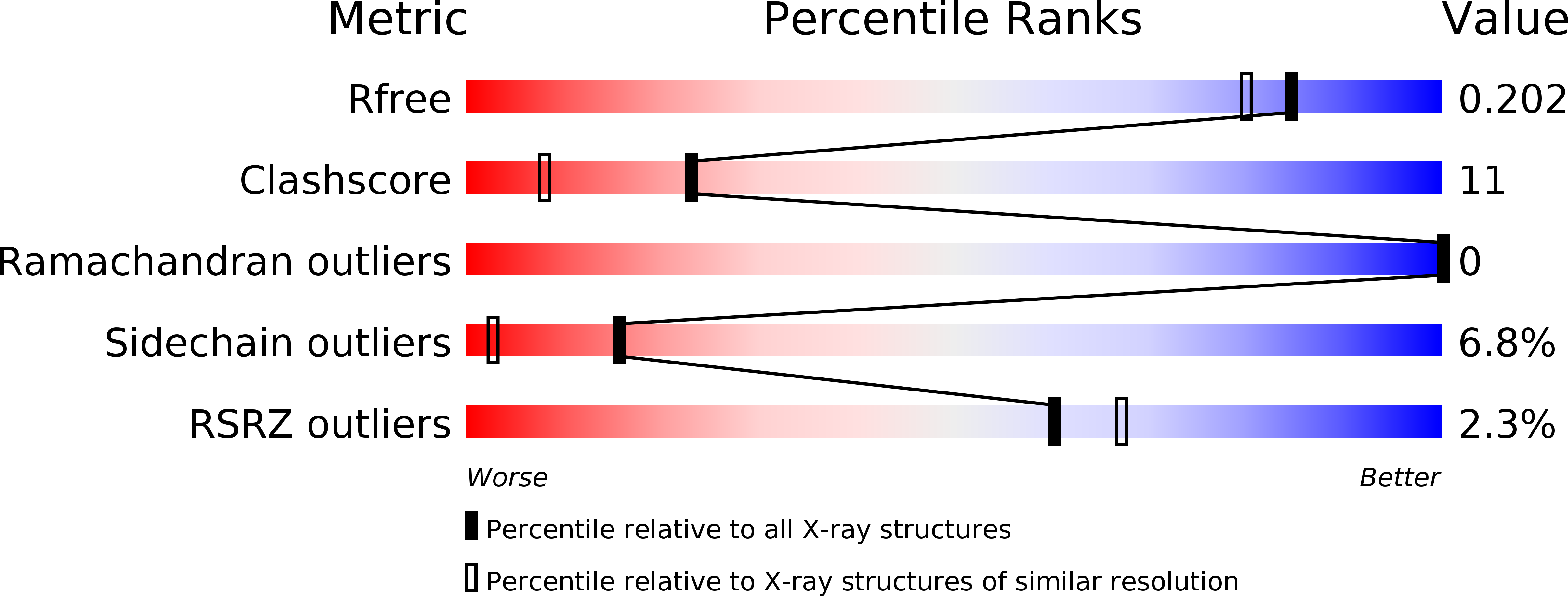
R-Value Free:
0.20
R-Value Work:
0.17
R-Value Observed:
0.17
Space Group:
H 3