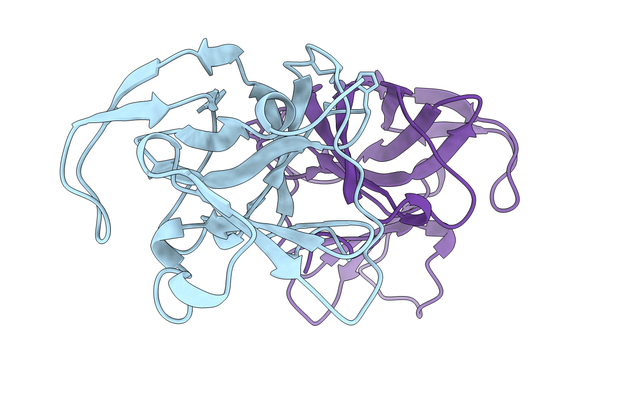
2HRV image
Deposition Date 1999-04-29
Release Date 2000-05-03
Last Version Date 2023-12-27
Entry Detail
PDB ID:
2HRV
Keywords:
Title:
2A CYSTEINE PROTEINASE FROM HUMAN RHINOVIRUS 2
Biological Source:
Source Organism:
Host Organism:
Method Details:
Experimental Method:
Resolution:
1.95 Å
R-Value Free:
0.25
R-Value Work:
0.21
R-Value Observed:
0.21
Space Group:
P 32 2 1
Macromolecular Entities
Structures with similar UniProt ID
Protein Blast
Polymer Type:polypeptide(L)
Molecule:2A CYSTEINE PROTEINASE
Chain IDs:A, B
Chain Length:142
Number of Molecules:2
Biological Source:Human rhinovirus 2
Ligand Molecules
Primary Citation
The structure of the 2A proteinase from a common cold virus: a proteinase responsible for the shut-off of host-cell protein synthesis.
EMBO J. 18 5463 5475 (1999)
PMID: 10523291 DOI: 10.1093/emboj/18.20.5463

Abstact

The crystal structure of the 2A proteinase from human rhinovirus serotype 2 (HRV2-2A(pro)) has been solved to 1.95 A resolution. The structure has an unusual, although chymotrypsin-related, fold comprising a unique four-stranded beta sheet as the N-terminal domain and a six-stranded beta barrel as the C-terminal domain. A tightly bound zinc ion, essential for the stability of HRV2-2A(pro), is tetrahedrally coordinated by three cysteine sulfurs and one histidine nitrogen. The active site consists of a catalytic triad formed by His18, Asp35 and Cys106. Asp35 is additionally involved in an extensive hydrogen-bonding network. Modelling studies reveal a substrate-induced fit that explains the specificity of the subsites S4, S2, S1 and S1'. The structure of HRV2-2A(pro) suggests the mechanism of the cis cleavage and its release from the polyprotein.

Legend

Protein

Chemical

Disease

Primary Citation of related structures