2HQV image
Deposition Date 2006-07-19
Release Date 2006-09-19
Last Version Date 2024-10-30
Entry Detail
PDB ID:
2HQV
Title:
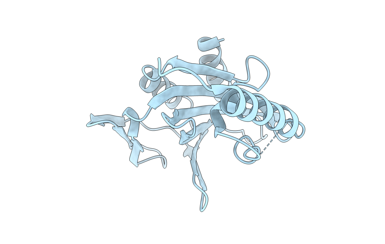
X-ray Crystal Structure of Protein AGR_C_4470 from Agrobacterium tumefaciens. Northeast Structural Genomics Consortium Target AtR92.
Biological Source:
Source Organism:
Host Organism:
Method Details:
Experimental Method:
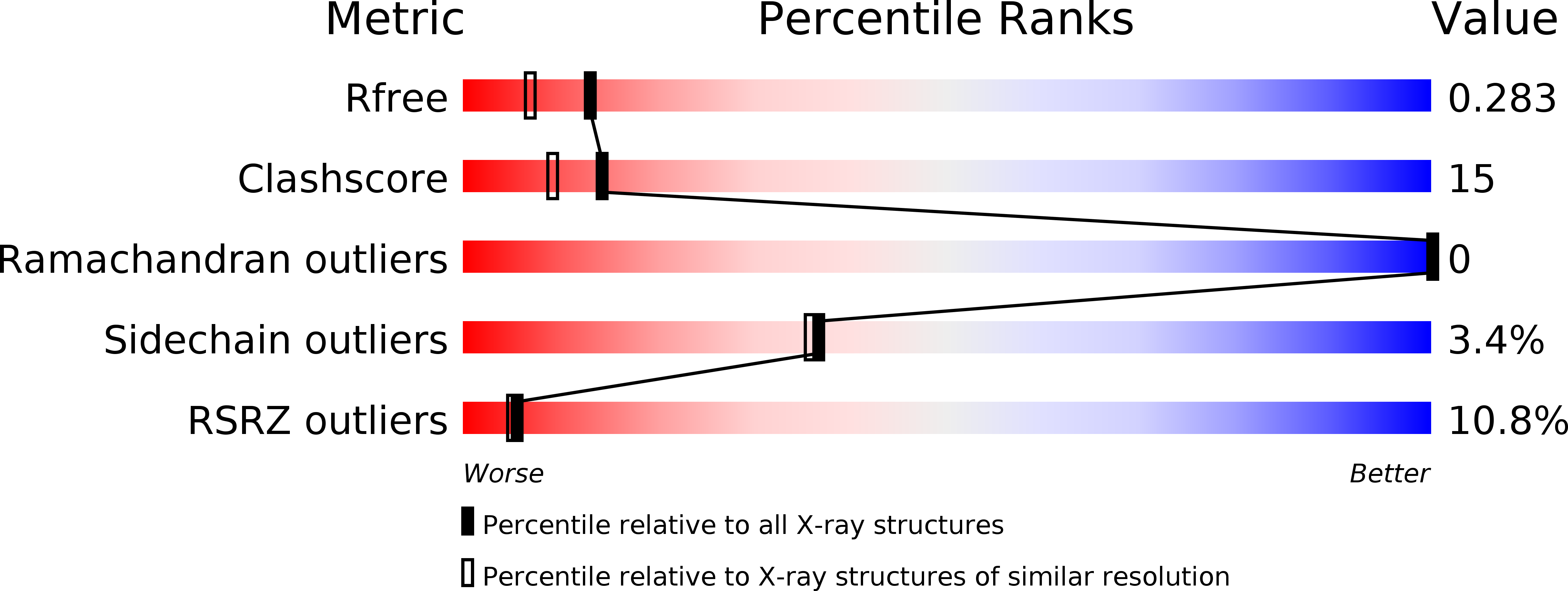
Resolution:
2.00 Å
R-Value Free:
0.26
R-Value Work:
0.24
R-Value Observed:
0.24
Space Group:
I 2 2 2
Macromolecular Entities
Polymer Type:polypeptide(L)
Molecule:AGR_C_4470p
Gene (Uniprot):Atu2462
Mutations:C-tag: LEHHHHHH
Chain IDs:A
Chain Length:195
Number of Molecules:1
Biological Source:Agrobacterium tumefaciens str. C58
Modified Residue
Compound ID Chain ID Parent Comp ID Details 2D Image
MSE A MET SELENOMETHIONINE
Primary Citation
Crystal structure of AGR_C_4470p from Agrobacterium tumefaciens.
Protein Sci. 16 535 538 (2007)
PMID: 17322535 DOI: 10.1110/ps.062663307

Abstact

We report here the crystal structure at 2.0 A resolution of the AGR_C_4470p protein from the Gram-negative bacterium Agrobacterium tumefaciens. The protein is a tightly associated dimer, each subunit of which bears strong structural homology with the two domains of the heme utilization protein ChuS from Escherichia coli and HemS from Yersinia enterocolitica. Remarkably, the organization of the AGR_C_4470p dimer is the same as that of the two domains in ChuS and HemS, providing structural evidence that these two proteins evolved by gene duplication. However, the binding site for heme, while conserved in HemS and ChuS, is not conserved in AGR_C_4470p, suggesting that it probably has a different function. This is supported by the presence of two homologs of AGR_C_4470p in E. coli, in addition to the ChuS protein.

Legend

Protein

Chemical

Disease

Primary Citation of related structures