Deposition Date
2006-07-17
Release Date
2006-08-08
Last Version Date
2024-02-14
Entry Detail
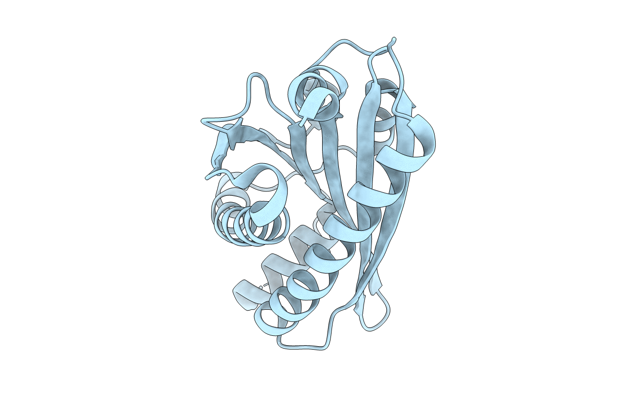
PDB ID:
2HP7
Keywords:
Title:
Structure of FliM provides insight into assembly of the switch complex in the bacterial flagella motor
Biological Source:
Source Organism(s):
Thermotoga maritima (Taxon ID: 2336)
Expression System(s):
Method Details:
Experimental Method:
Resolution:
2.00 Å
R-Value Free:
0.24
R-Value Work:
0.22
R-Value Observed:
0.22
Space Group:
P 43 21 2