
Deposition Date
2006-07-13
Release Date
2007-07-17
Last Version Date
2023-08-30
Entry Detail
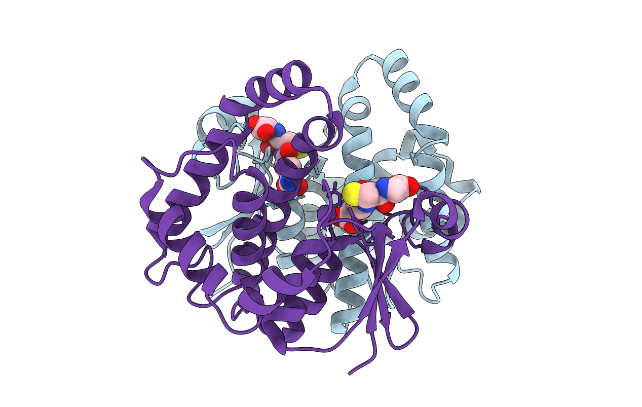
PDB ID:
2HNL
Keywords:
Title:
Structure of the prostaglandin D synthase from the parasitic nematode Onchocerca volvulus
Biological Source:
Source Organism(s):
Onchocerca volvulus (Taxon ID: 6282)
Expression System(s):
Method Details:
Experimental Method:
Resolution:
2.00 Å
R-Value Free:
0.23
R-Value Work:
0.18
R-Value Observed:
0.18
Space Group:
P 21 21 21
