
Deposition Date
2006-07-12
Release Date
2006-09-19
Last Version Date
2024-02-14
Entry Detail
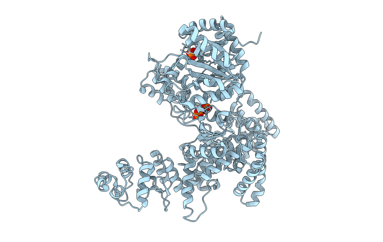
PDB ID:
2HNH
Keywords:
Title:
Crystal structure of the catalytic alpha subunit of E. coli replicative DNA polymerase III
Biological Source:
Source Organism(s):
Escherichia coli (Taxon ID: 562)
Expression System(s):
Method Details:
Experimental Method:
Resolution:
2.30 Å
R-Value Free:
0.25
R-Value Work:
0.18
R-Value Observed:
0.18
Space Group:
P 21 21 21
