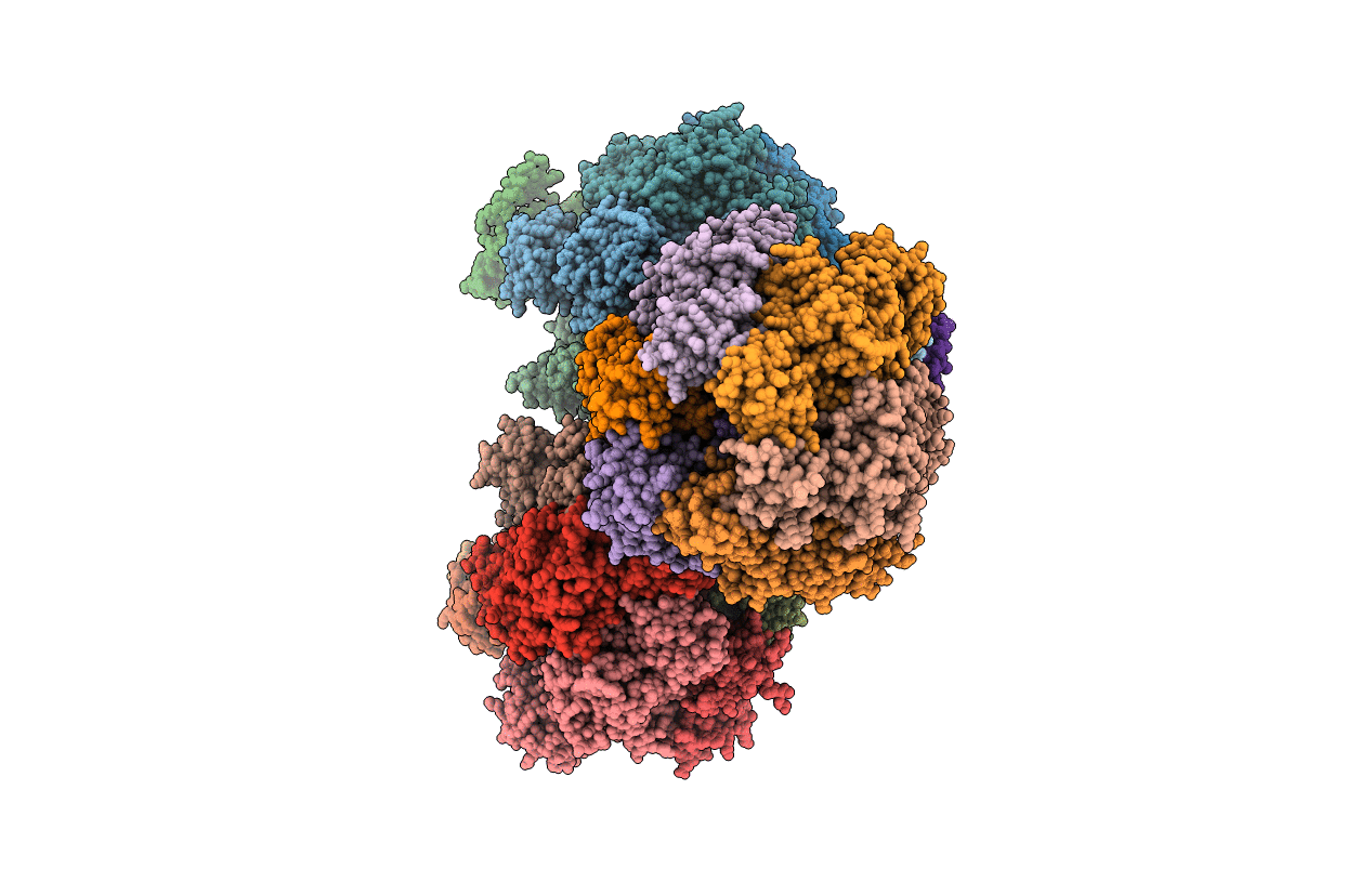
2HLD image
Deposition Date 2006-07-06
Release Date 2006-11-28
Last Version Date 2023-08-30
Entry Detail
PDB ID:
2HLD
Keywords:
Title:
Crystal structure of yeast mitochondrial F1-ATPase
Biological Source:
Source Organism:
Method Details:
Experimental Method:
Resolution:
2.80 Å
R-Value Free:
0.24
R-Value Work:
0.20
R-Value Observed:
0.20
Space Group:
P 1 21 1
Macromolecular Entities
Polymer Type:polypeptide(L)
Molecule:ATP synthase alpha chain, mitochondrial
Gene (Uniprot):ATP1
Chain IDs:A, B, C, J, K, L, S, T, U
Chain Length:510
Number of Molecules:9
Biological Source:Saccharomyces cerevisiae
Polymer Type:polypeptide(L)
Molecule:ATP synthase beta chain, mitochondrial
Gene (Uniprot):ATP2
Chain IDs:D, E, F, M, N, O, V, W, X
Chain Length:478
Number of Molecules:9
Biological Source:Saccharomyces cerevisiae
Polymer Type:polypeptide(L)
Molecule:ATP synthase gamma chain, mitochondrial
Gene (Uniprot):ATP3
Chain IDs:G, P, Y
Chain Length:278
Number of Molecules:3
Biological Source:Saccharomyces cerevisiae
Polymer Type:polypeptide(L)
Molecule:ATP synthase delta chain, mitochondrial
Gene (Uniprot):ATP16
Chain IDs:H, Q, Z
Chain Length:138
Number of Molecules:3
Biological Source:Saccharomyces cerevisiae
Polymer Type:polypeptide(L)
Molecule:ATP synthase epsilon chain, mitochondrial
Gene (Uniprot):ATP15
Chain IDs:I, R, AA (auth: 1)
Chain Length:61
Number of Molecules:3
Biological Source:Saccharomyces cerevisiae
Primary Citation

Abstact

The crystal structure of yeast mitochondrial F(1) ATPase contains three independent copies of the complex, two of which have similar conformations while the third differs in the position of the central stalk relative to the alpha(3)beta(3) sub-assembly. All three copies display very similar asymmetric features to those observed for the bovine enzyme, but the yeast F(1) ATPase structures provide novel information. In particular, the active site that binds ADP in bovine F(1) ATPase has an ATP analog bound and therefore this structure does not represent the ADP-inhibited form. In addition, one of the complexes binds phosphate in the nucleotide-free catalytic site, and comparison with other structures provides a picture of the movement of the phosphate group during initial binding and subsequent catalysis. The shifts in position of the central stalk between two of the three copies of yeast F(1) ATPase and when these structures are compared to those of the bovine enzyme give new insight into the conformational changes that take place during rotational catalysis.

Legend

Protein

Chemical

Disease

Primary Citation of related structures