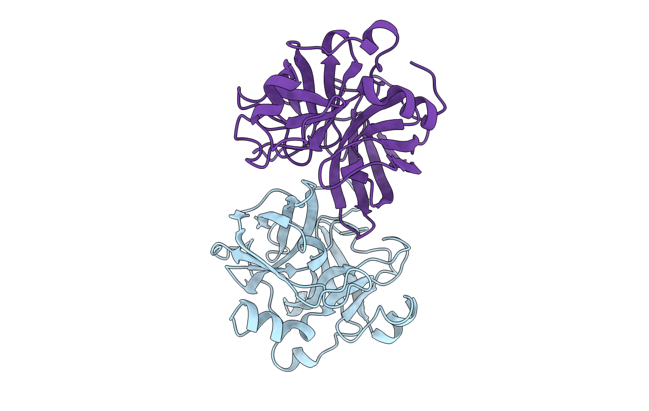
2HLC image
Deposition Date 1997-01-08
Release Date 1997-09-04
Last Version Date 2024-10-23
Entry Detail
PDB ID:
2HLC
Keywords:
Title:
HL COLLAGENASE STRUCTURE AT 1.7A RESOLUTION
Biological Source:
Source Organism:
Method Details:
Experimental Method:
Resolution:
1.70 Å
R-Value Free:
0.24
R-Value Work:
0.19
R-Value Observed:
0.19
Space Group:
I 4 2 2
Macromolecular Entities
Polymer Type:polypeptide(L)
Molecule:COLLAGENASE
Chain IDs:A, B
Chain Length:230
Number of Molecules:2
Biological Source:Hypoderma lineatum
Primary Citation
1.8 A structure of Hypoderma lineatum collagenase: a member of the serine proteinase family.
Acta Crystallogr.,Sect.D 52 380 392 (1996)
PMID: 15299709 DOI: 10.1107/S090744499501184X

Abstact

Collagenase from the fly larvae Hypoderma lineatum cleaves triple-helical collagen in a single region. It was crystallized at neutral pH in the absence of inhibitor and 1.8 A data were collected using synchrotron radiation and a Mark II prototype detector. The structure was solved by combining multiple isomorphous replacement methods and rotation translation function in real space. Refinement between 7 and 1.8 A using the program X-PLOR led to a final R factor of 16.9%. The overall fold is similar to that of other trypsin-like enzymes but the structure differs mainly by the presence of a beta-sheet at position 31-44. The two embedded molecules of the asymmetric unit are related by a pseudo twofold axis. The beta-sheet 31-44 of one molecule is involved in hydrogen bonds with binding-pocket residues of the other molecule. It thus completely prevents access to the active site. The specificity of this enzyme probably results from the position of Phe192 and Tyr99 at the entrance of the active site.

Legend

Protein

Chemical

Disease

Primary Citation of related structures