Deposition Date
2006-07-02
Release Date
2006-12-12
Last Version Date
2023-08-30
Entry Detail
PDB ID:
2HJV
Keywords:
Title:
Structure of the second domain (residues 207-368) of the Bacillus subtilis YxiN protein
Biological Source:
Source Organism(s):
Bacillus subtilis (Taxon ID: 1423)
Expression System(s):
Method Details:
Experimental Method:
Resolution:
1.95 Å
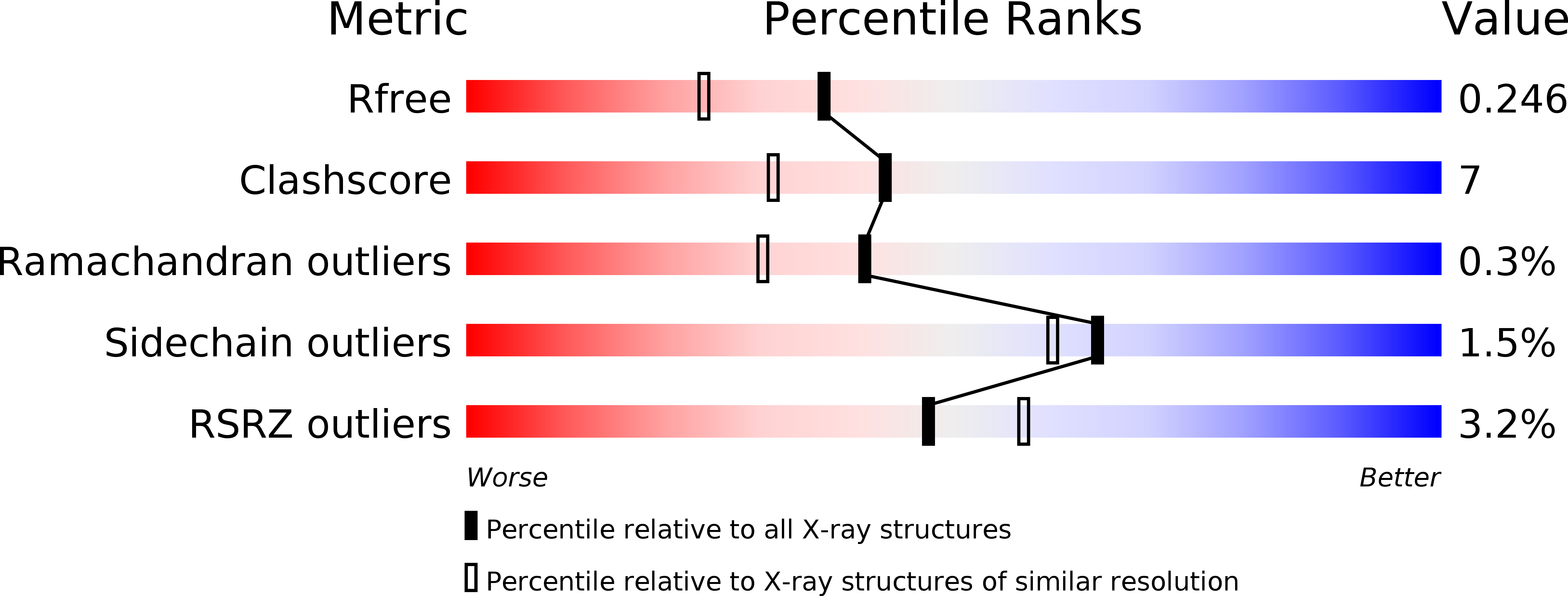
R-Value Free:
0.24
R-Value Work:
0.20
R-Value Observed:
0.20
Space Group:
P 61