2HJG image
Deposition Date 2006-06-30
Release Date 2006-08-08
Last Version Date 2024-11-20
Entry Detail
PDB ID:
2HJG
Keywords:
Title:
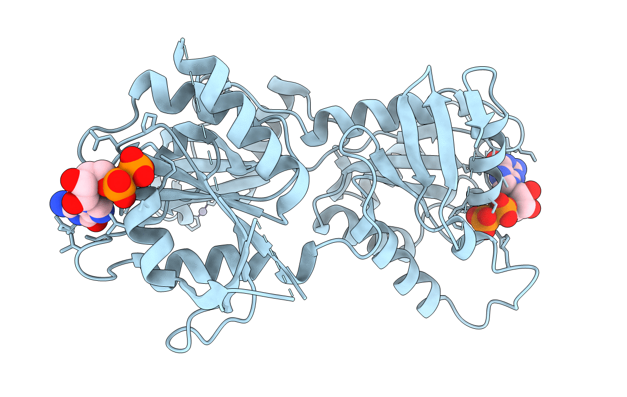
The crystal structure of the B. subtilis YphC GTPase in complex with GDP
Biological Source:
Source Organism:
Host Organism:
Method Details:
Experimental Method:
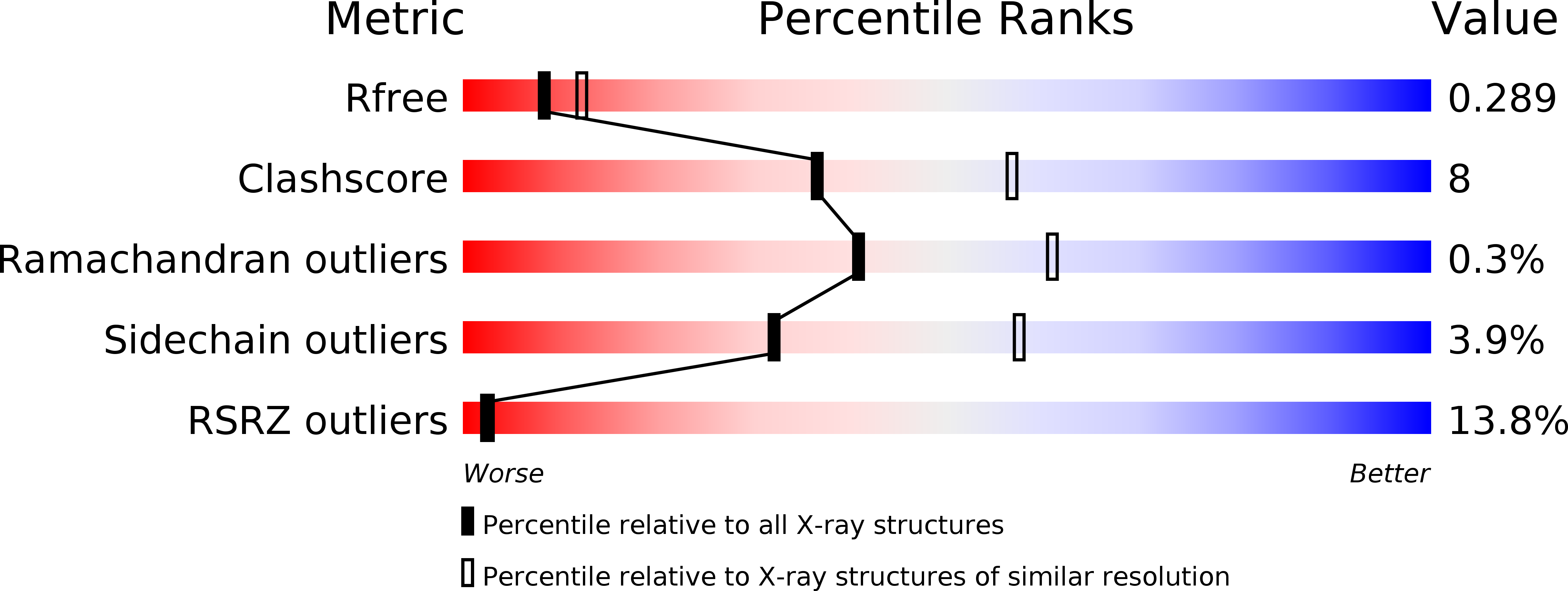
Resolution:
2.50 Å
R-Value Free:
0.27
R-Value Work:
0.21
R-Value Observed:
0.21
Space Group:
P 21 21 21
Macromolecular Entities
Structures with similar UniProt ID
Protein Blast
Polymer Type:polypeptide(L)
Molecule:GTP-binding protein engA
Gene (Uniprot):der
Chain IDs:A
Chain Length:436
Number of Molecules:1
Biological Source:Bacillus subtilis
Modified Residue
Compound ID Chain ID Parent Comp ID Details 2D Image
MSE A MET SELENOMETHIONINE
Primary Citation
The essential GTPase YphC displays a major domain rearrangement associated with nucleotide binding.
Proc.Natl.Acad.Sci.Usa 103 12359 12364 (2006)
PMID: 16894162 DOI: 10.1073/pnas.0602585103

Abstact

The structure of a Bacillus subtilis YphC/GDP complex shows that it contains two GTPase domains that pack against a central domain whose fold resembles that of an RNA binding KH-domain. Comparisons of this structure to that of a homologue in Thermotoga maritima reveals a dramatic rearrangement in the position of the N-terminal GTPase domain with a shift of up to 60 A and the formation of a totally different interface to the central domain. This rearrangement appears to be triggered by conformational changes of the switch II region in this domain in response to nucleotide binding. Modeling studies suggest that this motion represents transitions between the "on" and "off" states of the GTPase, the effect of which is to alternately expose and bury a positively charged face of the central domain that we suggest is involved in RNA recognition as part of the possible role of this enzyme in ribosome binding.

Legend

Protein

Chemical

Disease

Primary Citation of related structures